

Streamlined Western Blots with Stain-Free Precision
Achieve Accurate, Reproducible Results in Less Time with Innovative Stain-Free Technology
Stain-Free Technology
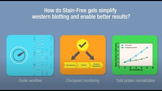
Stain-Free technology is superior to conventional western blotting methods as it allows users to:
- Visualize gels minutes after electrophoresis
- Perform quality assessments at each step without the need for additional staining and destaining
- Increase accuracy and produce reliable quantitative results via total protein normalization
RESOURCE SPOTLIGHT

Generate Western Blot Results You Can Stand Behind
Western blot data faces increasing scrutiny. Are your images fully defensible? This practical guide outlines how to document your images, apply consistent processing, and select the right normalization strategy to help you generate reproducible data that stands up to peer review.
Stain-Free Western Blotting Workflow


Separate Proteins
Speed with flexibility: TGX Stain-Free Gel chemistry available in precast and handcast formats

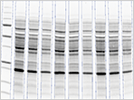
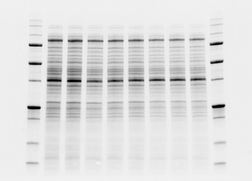
Pre-transfer gel image

Visualize Protein Separation
Coomassie-like performance with no background variability and no staining/destaining
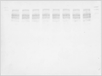
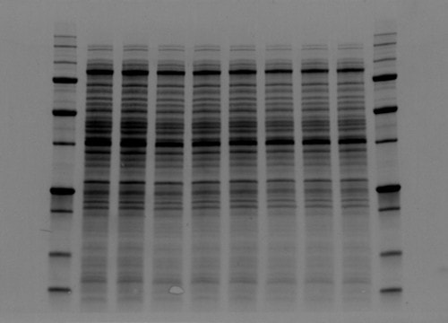
Post-transfer gel image

Protein Transfer
- Fast, efficient, and uniform protein transfer
- Throughput: transfer 4 mini or 2 midi gels at once
- Convenience: choose from ready-to-use transfer packs (nitrocellulose or PVDF)
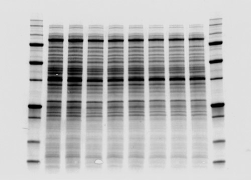
Blot gel image
Assess Transfer Efficiency
Quickly verify quality of transfer by imaging gel post transfer
Faster Blocking, Antibody Incubation, & Blot Detection Workflow:
A workflow that enables five-minute blocking, antibody incubation, and blot detection within approximately 4 hours — faster than traditional methods!
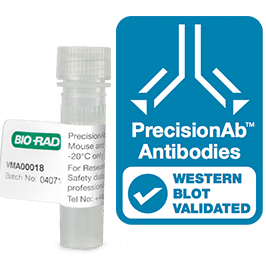
PrecisionAb Validated Western Blotting Antibodies
Highly specific and sensitive primary antibodies that have been extensively validated for western blotting.
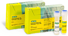
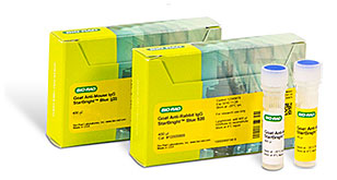
StarBright Blue Fluorescent Secondary Antibody
Ultra-sensitive fluorescence detection with very low background of single or multiple proteins in one blot without stripping or reprobing.
Normalize Total Protein and Analyze Western Blot Data
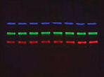
Easy multiplexing
Use western blot validated, highly specific PrecisionAb Primary Antibodies and our StarBright Blue Fluorescent Secondary Antibodies to detect multiple targets on the same blot.
-

Total protein detection with chemiluminescence
-
Total protein detection with fluorescence
-
Stain-free blot (from step 4) for normalization
Benefits of using Stain-Free blot image as total protein loading control
- Eliminates the need to strip then reprobe, and cut the blot
- More precise normalization methods compared to traditional methods which use a single housekeeping protein
- Produces reliable and accurate quantitation
Total Protein Normalization
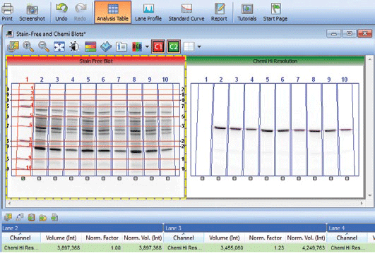
An illustration of the total protein normalization feature in the Image Lab Software using a Stain-Free enabled imager.
Bio-Rad’s Stain-Free western blotting provides a practical, convenient, and reliable way to perform total protein normalization. Stain-Free labeled proteins can be visualized in gels 1 minute after gel electrophoresis. Stain-Free blot visualization and image acquisition require only a few seconds using a stain-free enabled imager. This technology makes it easy for researchers to adopt total protein loading control for western blotting.
Total protein normalization using Stain-Free technology
- Enables total protein in load sample to be measured
- Eliminates immuno-blotting interference associated with HKP
- Accounts for variations during loading, electrophoresis, and transfer
Why is Stain-Free Total Protein Normalization Superior over Other Methods?
The use of traditional total protein stains can hinder downstream visualization of a western blot, usually requiring repetitive staining and detaining steps which is one of the obstacles preventing wider adaptation of TPN. The unique characteristics of Stain-Free technology makes imaging of total protein a much better method for quantitative western blotting.
-
No UV
-
With UV

Following a brief UV light activation, Stain-Free fluorochromes are covalently bound to protein molecules in the gel, allowing them to be imaged repeatedly on the gel or on a membrane post-transfer.
Total Protein normalization using Stain-Free technology consistently delivers scalar linear response across a wider dynamic range.
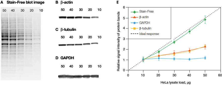
Linearity comparison of stain-free total protein measurement and immunodetection of three housekeeping proteins in 10–50 μg of HeLa cell lysate. On the left are representative images of (a), stain-free blot and the chemi blots for (b), β-actin; (c), β-tubulin and (d), GAPDH. Lane labels correspond to total protein load (μg). Although the actin and tubulin signals appear linear, the densitometric ratio was far below the predicted “quantitative response” of actual loading whereas the stain-free signal correlated to the expected result (e).
Reference
Taylor SC Posch A (2014). The design of a quantitative western blot experiment. Biomed Res Int. 36, 1590
Stain-Free technology allows scientists to visualize strong total protein signals with low background on their gels and blots that would otherwise not be possible without additional staining steps thus, improving overall dynamic range without the need to perform extra staining and destaining steps.
-
With Stain-Free Technology
-
Without Stain-Free Technology
Comparison between high background and low background signal in gels. Stain-Free technology noticeably lowers the background signal in comparison to traditional total protein detection methods.
Western Blotting Tips

Western Blot Selector Tool
Get the best reagents and consumables to ensure optimized western blotting performance.
Sign Up to Be the First to Know
Receive Western Blotting Tips and Online Resource Updates
Keep up to date with useful tips to continuously improve your western blotting experiments from sample preparation through image analysis. Sign up to be the first to be notified when new western blotting resources like tips and tricks, posters, protocols, webinars and how-to-videos become available.
Ordering
Devices
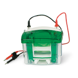
Mini-PROTEAN Tetra Vertical Electrophoresis Cell for Mini Precast Gels, 4-gel #1658004
4-gel vertical electrophoresis system, includes electrode assembly, companion running module, tank, lid with power cables, mini cell buffer dam

Criterion Cell #1656001
Vertical midi-format electrophoresis cell, includes buffer tank, lid with power cables, 3 sample loading guides (12+2-well, 18-well, and 26-well)
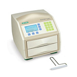
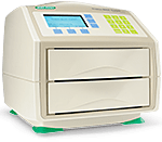
Trans-Blot Turbo Transfer System #1704150
Blotting instrument, includes base, 2 cassettes to hold 1–2 midi or up to 4 mini blotting sandwiches, blot roller
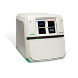
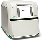
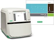
ChemiDoc Imaging System #12003153
The ChemiDoc Imaging System allows sensitive imaging and documentation of DNA/protein gels and chemiluminescent western blots

ChemiDoc MP Imaging System #12003154
The ChemiDoc MP Imaging System is a full-feature, best-in-class instrument for imaging and quantifying nucleic acid and protein in gels and western blots. It is capable of imaging fluorescent western blots (RGB, far-red, near-IR), chemiluminescent western blots, all common nucleic acid and protein gel stains, plus stain-free gels. Includes internal computer, 12" touch-screen display, camera, Image Lab Touch Software, and Image Lab Software

ChemiDoc Go Imaging System with Image Lab Touch Software #12018025
Modern, compact imaging system with advanced chemiluminescence and StarBright™ Blue fluorescence detection and BR.io cloud connectivity. The ChemiDoc Go Imaging System allows sensitive imaging and documentation of DNA gels, protein gels, and chemiluminescent western blots. With an advanced CMOS digital detector, all-LED light sources for trans- and epi-illumination, and compatibility with Stain-Free Western Blotting.
Kits and Reagents
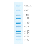
Precision Plus Protein All Blue Prestained Protein Standards #1610373
500 µl, mixture of ten blue-stained recombinant proteins (10–250 kD), including three reference bands (25, 50, 75 kD), 50 applications
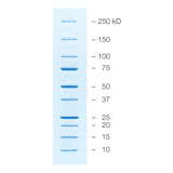
Precision Plus Protein Unstained Protein Standards, Strep-tagged recombinant, 1 ml #1610363
Precision Plus Protein Unstained Protein Standards, Strep-tagged recombinant, 1 ml>1 ml, unstained mixture of ten Strep-tagged, recombinant proteins (10–250 kD), including three reference bands (25, 50, 75 kD), 100 applications
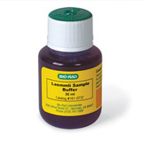
2x Laemmli Sample Buffer #610737
30 ml, premixed protein sample buffer for SDS-PAGE
10x Tris/Glycine/SDS #1610732
Pkg of 1, 1 L, 10x premixed electrophoresis buffer, contains 25 mM Tris, 192 mM glycine, 0.1% SDS, pH 8.3 following dilution to 1x with water

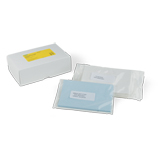
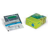
Trans-Blot Turbo Mini 0.2 µm PVDF Transfer Packs #1704156
Pkg of 10, 7 x 8.5 cm, precut blotting transfer pack, includes filter paper, buffer, 0.2 µm PVDF membrane, for use with Trans-Blot Turbo Transfer System
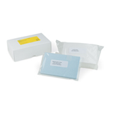
Trans-Blot Turbo Midi 0.2 µm PVDF Transfer Packs #1704157
Pkg of 10, 8.5 x 13.5 cm, precut blotting transfer pack, includes filter paper, buffer, 0.2 µm PVDF membrane, for use with Trans-Blot Turbo Transfer System

Trans-Blot Turbo Mini 0.2 µm Nitrocellulose Transfer Packs #1704158
Pkg of 10, 7 x 8.5 cm, precut blotting transfer pack, includes filter paper, buffer, 0.2 µm nitrocellulose membrane, for use with Trans-Blot Turbo Transfer System
Trans-Blot Turbo Midi 0.2 µm Nitrocellulose Transfer Packs #1704159
Pkg of 10, 8.5 x 13.5 cm, precut blotting transfer pack, includes filter paper, buffer, 0.2 µm nitrocellulose membrane, for use with Trans-Blot Turbo Transfer System
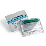
Immun-Blot Low Fluorescence PVDF/Filter Paper Sets #1620260
Pkg of 10, 0.45 µm, 7 x 8.5 cm, precut low fluorescence PVDF/filter paper for immunoblotting

Immun-Blot Low Fluorescence PVDF/Filter Paper Sets #1620261
Pkg of 20, 0.45 µm, 7 x 8.5 cm, precut low fluorescence PVDF/filter paper for immunoblotting
Immun-Blot Low Fluorescence PVDF/Filter Paper Sets #1620262
Pkg of 10, 0.45 µm, 8.5 x 13.5 cm, precut low fluorescence PVDF/filter paper for immunoblotting
Immun-Blot Low Fluorescence PVDF/Filter Paper Sets #1620263
Pkg of 20, 0.45 µm, 8.5 x 13.5 cm, precut low fluorescence PVDF/filter paper for immunoblotting

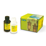
EveryBlot Blocking Buffer, 500 ml #12010020
Bottle of blocking buffer, 500 ml. Requires only 5 minutes of blocking for all western blots and eliminates incubation time in ELISA assays.
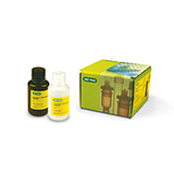
Clarity Western ECL Substrate, 200 ml #1705060
Pkg of 1, contains 100 ml Clarity Western Peroxide Reagent and 100 ml Clarity Western Luminol/Enhancer Reagent
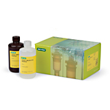
Clarity Western ECL Substrate, 500 ml #1705061
Pkg of 1, contains 250 ml Clarity Western Peroxide Reagent and 250 ml Clarity Western Luminol/Enhancer Reagent
Clarity Max Western ECL Substrate, 100 ml #1705062
Pkg of 1, contains 50 ml Clarity Max Western Peroxide Reagent and 50 ml Clarity Max Luminol/Enhancer Reagent
Gels
Mini-PROTEAN TGX Stain-Free Precast Gels
Based on the long-shelf-life TGX (Tris-Glycine eXtended) formulation and include unique trihalo compounds that allow rapid fluorescent detection of proteins with the ChemiDoc MP and GelDoc Go Imaging Systems.
Criterion TGX Stain-Free Precast Gels
Based on the long shelf life TGX formulation and include unique trihalo compounds that allow rapid fluorescent detection of proteins with the ChemiDoc MP and GelDoc Go Imaging Systems.
Antibodies
-
PrecisionAb Validated Western Blotting Antibodies
Collection of highly specific and sensitive primary antibodies (monoclonal and polyclonal) that have been extensively validated for western blotting.
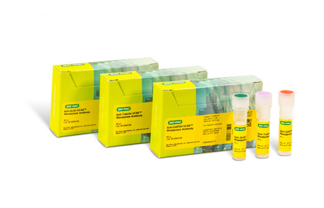
hFAB Rhodamine Housekeeping Protein Fluorescent Primary Antibodies
One-step detection of common housekeeping proteins like actin, tubulin, and GAPDH in your samples. These anti-actin, anti-tubulin, and anti-GAPDH antibodies are directly labeled with a rhodamine derivative (Exmax/Emmax = 530 nm/580 nm).
StarBright Blue Fluorescent Secondary Antibody
Conjugated to a high-yield fluorophore, for ultra-sensitive fluorescence detection with very low background of single or multiple proteins in one blot without stripping or reprobing.
Resources

Stain-Free Western Blotting with ChemiDoc MP Imaging System

Stain-Free Approach for Western Blotting
(PDF 885 KB)
A Defined Methodology for Reliable Quantification of Western Blot Data
(PDF 602 KB)-
Trends in Protein Separation and Analysis — the Advance of Stain-Free Technology

Western Blot Normalization Using Image Lab Software
(PDF 444 KB)
Determining the Appropriate Sample Load When Using a Stain-Free Western Workflow
(182 KB)
Validating the Expression Consistency of a Housekeeping Protein
(198 KB)
Western Blotting Protocol Library
Filter by your laboratory set-up and reagents to get a custom western blotting protocol that best fits your needs.-
Image

Western Blot University
Courses designed to make you a western blotting expert.


Stain-Free Western Blotting Resource Collection
Four-part literature series created to help researchers effectively learn and apply Stain-Free Western Blotting techniques. It provides comprehensive guidance on mastering both Stain-Free workflows and Total Protein Normalization.

The Complete Guide to Stain-Free Western Blotting
Download this free eBook to learn how incorporating Stain-Free technology can revolutionize your western blotting.
Applications & Technologies

Stain-Free Imaging Technology
Visualize proteins on gels and blots without staining and destaining steps, and use total protein normalization for quantitative western blots.

Western Blotting Techniques
Learn more about western blotting techniques. Find step-by-step protocols and helpful tips on equipment, membranes, transfer conditions, and detection methods.

Protein Separation and Analysis
Bio-Rad's Stain-Free Western Blotting Workflow facilitates speed and validation at each step of a western blotting experiment — from running gels to quantifying proteins.

Protein Electrophoresis
Find protocols, video tutorials, and selection guides to help you at every step of your electrophoresis experiments.

Imaging and Analysis
Find information on protein visualization and quantitation methods, gel and blot imaging instrumentation, and image analysis software.


