Introduction to In-Cell Western Assays
In-Cell Western (ICW) is a powerful quantitative and qualitative technique that enables the analysis of protein expression in-situ without the hassle of cell lysate sample preparation.
-
The In-Cell Western User:
In-Cell Western is ideal for those who have already validated the specificity of their antibodies with traditional western blotting or for those interested in overall protein expression without needing to account for molecular weight.
-
Uses for In-Cell Western:
- Quantification of protein expression changes in response to drug treatments or genetic manipulations
- Evaluation of protein-proteininteractions
- Screening large libraries of compounds in drug discovery and development
- Analysis of protein localization within specific cellular compartments
Example of a typical In-Cell Western workflow

In-Cell Western Protocol vs Traditional Western Blotting (Total Protocol Length)
The In-Cell Western Assay is an efficient, time-saving method ideal for those who have already validated the specificity of their antibodies with traditional western blotting or for those interested in overall protein expression without needing to account for molecular weight.
-
Total Time for In-Cell Western (up to 384 samples)
-
Total Time for Western Blot (up to 24 samples)
Advantages of In-Cell Western Assay vs. Traditional Western Blotting
-
Advantage 1:
Shorter protocol — no need for time-consuming sample preparation, electrophoresis, and transfer steps.
-
Advantage 2:
Quantification of multiple targets (up to 3-plex) in numerous samples (up to 384-wells).
-
Advantage 3:
Protein detection in situ — qualitative and quantitative immunofluorescence.
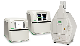
In-Cell Western Assays Made Easy Using the ChemiDoc MP System
The top reasons why our ChemiDoc MP System is superior to competitor imagers for In-Cell Western Imaging:
- Enables multiplexing of up to 3 targets — unlike methods from competitors that only allow up to 2 targets
- Not constrained to NIR dyes — ChemiDoc MP System has five fluorescent channels
- Provides superior image quality and pairs seamlessly with Image Lab Software for simplistic analysis
- Equipped with ready-to-use controls when combined with our hFAB Rhodamine Housekeeping Protein Fluorescent Primary Antibodies
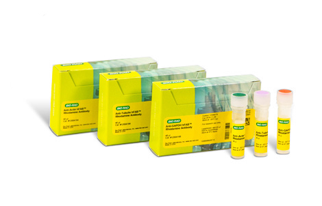
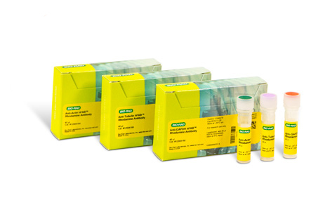
Find the Right hFAB Rhodamine Housekeeping Protein Fluorescent Primary Antibody for Your Experiment
Bio-Rad offers commonly used house-keeping primary antibodies conjugated to a rhodamine dye, making it easy to add to any assay and a natural control.
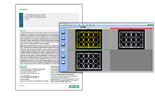
Image Lab Analysis of In-Cell Western Assays
Image Lab Software simplifies In-Cell Western analysis by taking advantage of its harmonious pairing with any ChemiDoc Imager, making it a powerful yet easy-to-use package for acquisition and analysis of your In-Cell Western Plate. By using the downloadable plate templates provided on this page or creating your own using the Volume Tool, Image Lab Software automatically calculates volume intensity of each well. When coupled with Bio-Rad’s hFAB Rhodamine Housekeeping Protein Fluorescent Primary Antibodies, you can set control wells along with blank wells that are used for background subtraction to calculate adjusted volume intensity. In-Cell Western analysis allows for quantification of multiple targets using spectrally distinct fluorescent dyes and is a quick method for finding relative protein levels in numerous samples.
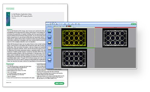
Templates and Protocol
Download these pre-made plate templates and optimized protocol to simplify your In-Cell Western Image Lab Analysis:
Get Templates and ProtocolApplications & Technologies
-
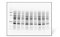
Western Blotting
Learn about the powerful technique that allows you to positively detect your proteins, estimate quantities, and determine their molecular weights.
-
Western Blot Learning Center
The Western Blot Learning Center is a complete reference on all of the steps of western blotting, includes practical theory, protocols, and recommendations on how to make your blots better from experts.
Related Products
-
ChemiDoc Imaging Systems
See our stain-free imaging systems that enable immediate visualization of proteins without gel staining and instant verification of protein transfer to blots.
-
Image Lab Analysis Software
View the Image Lab Software, an intuitive yet powerful analysis software that is compatible with ChemiDoc Imaging Systems and the GelDoc Go Imaging System.
-
hFAB™ Rhodamine HKP Antibodies
Select our hFAB Rhodamine Housekeeping Protein Primary Antibodies that allow one-step detection of common housekeeping proteins such as actin, tubulin, and GAPDH.

