Image

Description
Description
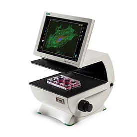
Use the ZOE Fluorescent Cell Imager for routine cell culture and imaging applications.
With brightfield and multichannel fluorescence, an integrated digital camera, and a touch-screen interface, this inverted microscope makes cell imaging simple, fast, and intuitive.
Features of the ZOE Cell Imager include:
- Simplified cell imaging — the intuitive touch-screen interface allows users to view cells, capture images, and create multichannel merges with minimal training
- Flexible operation — one brightfield and three fluorescent channels enable use for routine cell culture applications as well as more sophisticated imaging applications
- Fluorescence at your bench — light shield permits fluorescence imaging in ambient light
- Robust construction — fully integrated system with long-life LEDs, ready for intensive daily use
- LED light sources — thousands of hours of illumination that are instantly ready after power-on
- Large imaging area — the motorized stage and wide field of view allow you to see more of your sample, faster
- Small footprint — compact size accommodates crowded lab benches
Illumination light sources
- Blue channel uses a UV LED
- Green channel uses a blue LED
- Red channel uses a green LED
- Brightfield channel uses a ring of multiple green LEDs for reduced chromatic aberration
Fluorescence channels
- Blue channel: excitation, 355/40 nm; emission: 433/36 nm
- Green channel: excitation, 480/17 nm; emission: 517/23 nm
- Red channel: excitation, 556/20 nm; emission, 615/61 nm
The ZOE Fluorescent Cell Imager is compatible with many commonly used fluorescent dyes and proteins.
Specifications
Specifications
Imaging channels
Brightfield channel and 3 fluorescence channels (blue, green, and red)
Light source
Blue channel: UV LED
Green channel: blue LED
Red channel: green LED
Brightfield channel: multiple green LEDs (reduces chromatic aberration)
Green channel: blue LED
Red channel: green LED
Brightfield channel: multiple green LEDs (reduces chromatic aberration)
User interface
10.1 in. color (26 cm) touch-screen LCD monitor, with anti-glare and anti-fingerprint treatment, 1,280 x 768 pixel image resolution, 80–180° angle tilt range
Focusing mechanism
Coarse and fine, manual adjustment
Camera
Monochrome camera, 12 bit CMOS, 5 megapixels
Data format
JPEG, TIFF, or RAW image files
Image merge
Images from up to 4 channels can be overlaid
Data storage
16 GB internal memory (~ 2,500 JPEG files,1,500 TIFF files, 400–800 RAW files)
Data export
Yes, 2 USB ports
Display output
Yes, 1 HDMI port
Objective
20x
Numerical aperture
0.40
Display magnification
Standard: 175x; zoom: 700x
Maximum imaging area
0.70 mm2 field of view
Motorized stage
6 mm travel in X, Y direction, touch-screen control of travel speed and direction
Compatible with
Flasks: T25, T75, or T225
Multiwell plates: 6-, 12-, 24-, 48-, 96-, or 384-well microplates
Dishes: 35 mm, 60 mm, or 100 mm
Slides: chamber slides or standard glass microscopy slides
Multiwell plates: 6-, 12-, 24-, 48-, 96-, or 384-well microplates
Dishes: 35 mm, 60 mm, or 100 mm
Slides: chamber slides or standard glass microscopy slides
Software
Stand-alone Android operating system; PC is not required for operation
Instrument size (L x W x H)
33 x 32 x 30 cm (13 x 12.6 x 11.6 in.)
Instrument weight
9 kg (19.7 lb)
