
The Bio-Plex Suspension Array System utilizes xMAP technology licensed from Luminex to permit the multiplexing of up to 100 different assays within a single sample. This technique involves 100 distinctly colored bead sets created by the use of two fluorescent dyes at various ratios. In a sandwich immunoassay, one antibody to a specific analyte is attached to a set of beads with the same color, and the second antibody against the analyte is attached to a fluorescent dye. A dual detection flow cytometer is used to identify the different assays based on bead color in one channel, and to quantify the analyte by measurement of the reporter dye fluorescence in another channel.
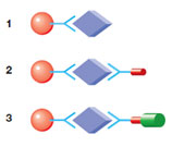
Bio-Plex Assay.
In a typical Bio-Plex Assay, a capture molecule conjugated to a color-coded bead binds to a target analyte (1) followed by binding with biotinylated detection antibody (2) and a reporter molecule, streptavidin-PE (3).
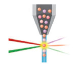
Bio-Plex Reader.
In the Bio-Plex Array Reader, a red classification and a green reporter laser illuminate individual beads to identify each bead's spectral address and associated reporter signal.
Related Topics: Bio-Plex Assays and Bio-Plex Assay Data Analysis.
-
FEATURED ARTICLEAberrant Cytokine Activity in the Host Immune
Response to COVID-19 Leads to Cytokine
Release Syndrome
Page Contents
After the analytes have been captured and coupled with the detection antibodies, the Bio-Plex Instrument or Array Reader is then used to detect the identity and determine the quantity of the analytes.
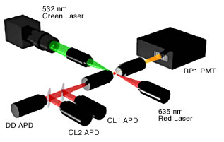
Components of the Bio-Plex Reader. APD, avalanche photodiode detector; DD, doublet discriminator channel, discriminates single beads from aggregated beads; CL1, classify channel, allows multiplexing, detects dye inside beads; and RP1, reporter channel, quantitates assay in this channel.
The Bio-Plex Array Reader comprises four different components:
- Fluidics — the Bio-Plex Array Reader detects individual beads by flow cytometry. The fluidics system of the reader aligns the beads into single file as they enter a stream of sheath fluid and then enter a flow cell. Once the beads are in single file within the flow cell, each bead is individually interrogated for bead color (analyte) and assay signal strength (phycoerythrin fluorescence intensity)
- Lasers — the reader is a dual laser system. The 532 nm Nd:YAG laser (green "reporter" laser) is used to excite the phycoerythrin (PE) dye of the assay, for example, streptavidin-PE used for sandwich immunoassays. The 635 nm solid state laser (red "classify" laser) is used to excite the dyes inside the beads to determine their "color" or "region" and is also used for doublet discrimination by light scatter
- Optics — the reader contains an optics bench with four detection channels. The dual lasers are precisely focused to excite an individual bead within the quartz flow cell. Upon excitation, the fluorescent signal emitted from the bead travels though the optics paths to the individual detectors
- Detectors — the reader has four detectors, one for each of the optical paths shown in the figure above. A high-sensitivity photomultiplier tube (PMT) detector is used for the reporter channel. Photodiodes are used for the stronger signals of the classify CL1 and CL2 and doublet discrimination channels
Related Content
Videos


Dr. Spinale, Professor of Cardiothoracic Surgery at Medical University of South Carolina, explains how the Bio-Plex System allowed him to tackle important questions in childhood disease research.