After 2-D gels are stained, the protein patterns are digitized and analyzed. Following analysis, spots of interest are then excised from gels for further analysis. Bio-Rad provides a wide assortment of imaging systems to meet various 2-D gel imaging requirements. Sophisticated image analysis software simplifies the analysis of 2-D data and makes the process more efficient by automating spot detection and analysis. This section provides an overview of gel image acquisition instruments and analysis software.
Related Topics: Protein Sample Preparation for 2-D Electrophoresis, First Dimension Separation (Isoelectric Focusing), Second Dimension Separation, Visualization (Staining), Protein Spot Excision and Protein Identification, and Troubleshooting 2-D Electrophoresis Gels with 2-D Doctor™.
Page Contents
Analysis of high-quality 2-D gels is a basic requirement for investigating changes in protein expression. Before 2-D gels can be analyzed with an image evaluation system, they must be digitized. The most commonly used devices are camera-based systems, densitometers, phosphor imagers, and fluorescence scanners. Bio-Rad has imaging equipment suitable for different kinds of detection methods.
Imaging Systems
 |
 |
 |
|
| GS-900™ LED illumination |
ChemiDoc™ MP | Gel Doc™ EZ | |
| Type of Imager | Densitometer | CCD camera-based | CCD camera-based |
| Light source options | Epl- and transillumination of white light | Transillumination of UV and white light with near infrared, far red, red, green, and blue LEDs | Transillumination of UV and white light |
| Optimized applications | |||
| Colorimetric stains | • | • | • |
| UV stains | — | • | • |
| Fluorescent stains and labels | — | • | — |
| Fluorescent multiplexing | — | • | — |
| Chemilluminescence | — | • | — |
| Stain-Free | — | • | • |
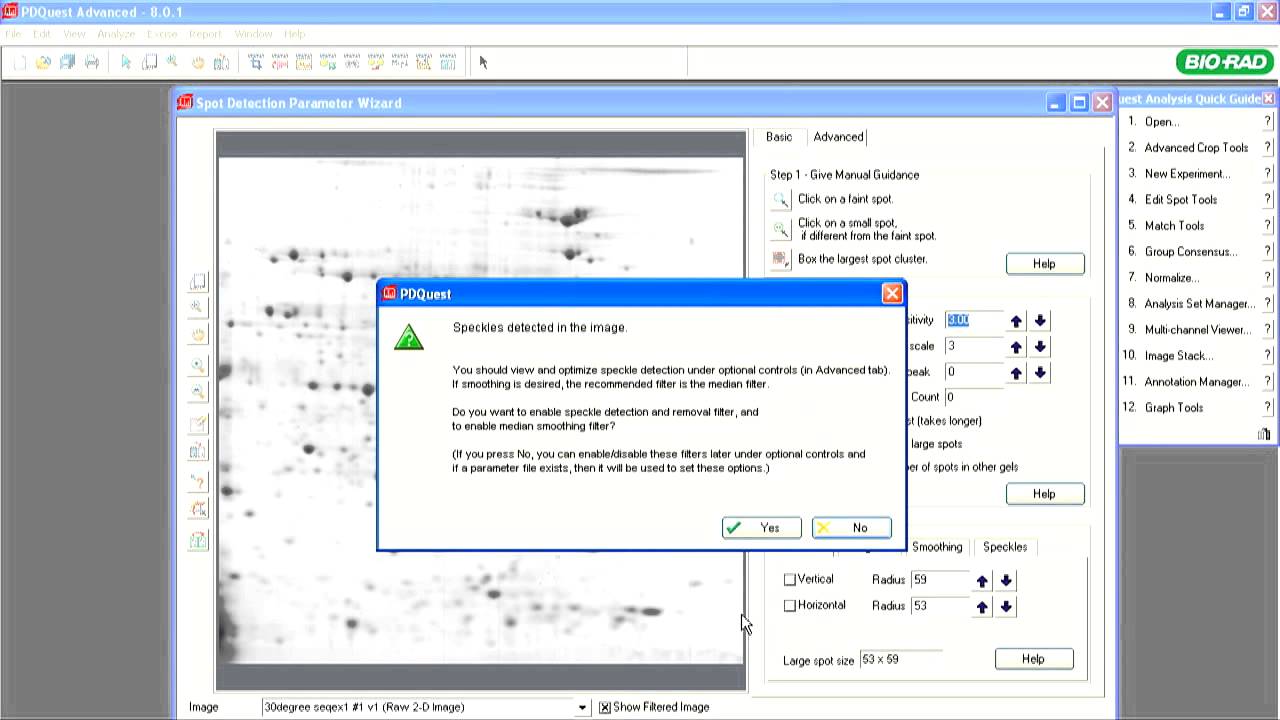
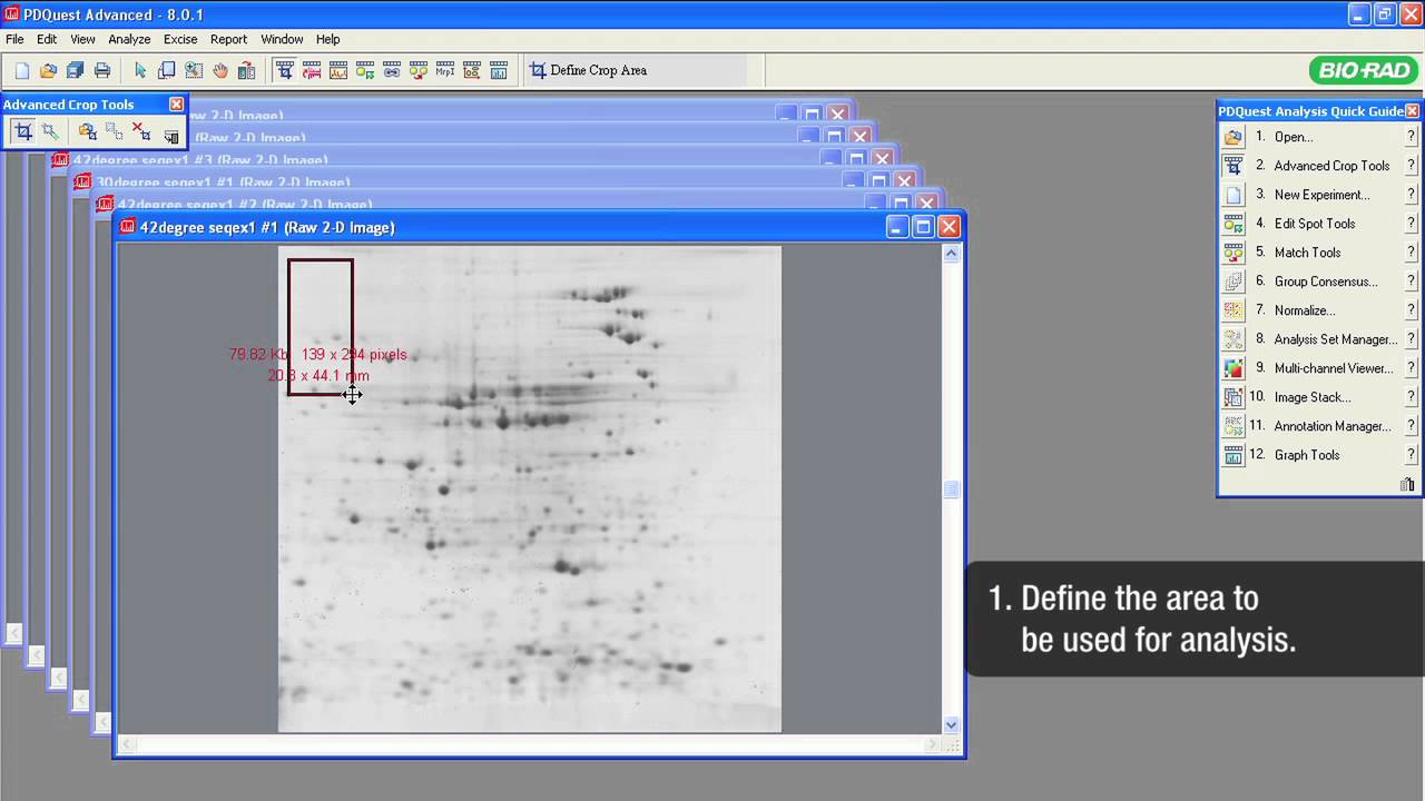
Computer-assisted image analysis is an indispensable tool for the evaluation of complex 2-D gels. Image analysis software obtains quantitative and qualitative information about the proteins in a sample and stores the information in files, which may also contain additional annotations. PDQuest™ 2-D analysis software is a tool for analysis of digitized gel images that integrates with Bio-Rad's image acquisition instruments.
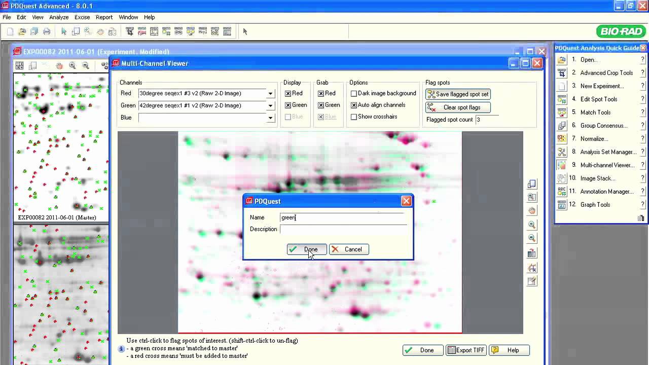
Spot comparison with PDQuest software. PDQuest software can be used to compare two or more gel images, revealing changes in protein expression in response to different treatments, growth conditions or disease states.
The wizard driven software uses the following work flow:

PDQuest can also be used to control the ExQuest™ spot cutter for automated spot excision.
After separation by 1-D gel electrophoresis, you can utilize a number of methods for protein imaging and analysis involving staining techniques or stain-free technology in combination with specialized imaging devices and accompanying software.