

Introduction
Transfection is the delivery of nucleic acids and other molecules into eukaryotic cells through nonviral methods while viral-based transfection or transduction involves using a viral vector to carry a specific nucleic acid sequence into a host cell (Chong ZX et al. 2021). The advancement of these technologies has enabled scientists to investigate protein function and gene regulation in a variety of cell types, tissues, and organisms. Gene delivery can be achieved by these three different methods: chemical, physical, and viral. Chemical transfection can be achieved via lipid-mediated approaches, whereas physical transfection methods could include electroporation, biolistic particle delivery, microinjection, mechanoporation, and other physical methods.


The optimal method for gene delivery depends on the specific application, delivery molecule(s), and the target cell type, all of which can influence the overall effectiveness of the gene approach used. Once gene delivery has been completed, various techniques are available for post-transfection or transduction analysis and for evaluating gene delivery efficiency as well as cell viability.
Another parameter to consider is the timeframe for the delivery process, and whether a transient or stable event is desired. Transient gene expression can be useful for evaluating gene promoter function. Stable expression leads to integration of the gene of interest into the host cell genome. This method is often used to study long-term effects of gene expression or to create cell lines with novel characteristics for further downstream studies, e.g., screening potential drug candidates in drug discovery experiments. Further information on different methods of gene delivery can be found on the Introduction to Transfection page.
Factors That Can Impact Gene Delivery Efficiency
Featured Content
Methods to Assess Gene Delivery Efficiency & Cell Viability
After a gene delivery event, it is important to assess delivery efficiency and the impact on cell health. This may be as simple as confirming the expression of a gene of interest. Alternatively, it may be important to quantify viability or the level of expression in some cases. This can be achieved using various methods including cell counting, flow cytometry, western blotting, imaging, real-time quantitative PCR (qPCR), and droplet digital PCR (ddPCR).
A typical gene delivery analysis process may include determining the percentage of positive cells within a transfected population, measuring the degree of expression in relative or absolute terms, and/or visual confirmation of a protein of interest and its localization. PCR-based approaches are well-suited for the accurate quantification of gene expression; however, they lack the ability to provide direct evidence of protein expression. In contrast, alternate methods such as flow cytometry and western blotting offer direct evidence of protein expression but with less precision.
Here we discuss general guidelines and methods for analyzing transfected or transduced cells and explore factors to consider when performing gene and protein expression analysis. While the utility of each of these techniques is well accepted, the most appropriate method in each case should be determined based on the nature of the investigation and resources available.
Cell Counting

Cell counting is a crucial technique for analyzing transfection efficiency, providing a quick and reliable method to determine the proportion of viable cells prior to or following gene delivery. This can be performed manually using a hemocytometer or with an automated cell counter (Gallicano GI et al. 2022). Automated cell counting systems have the advantage of allowing faster counting while at the same time facilitating standardization and reproducibility. Additionally, cell counting is an essential step inherent to many of the analysis techniques discussed in this guide and will often be needed multiple times throughout a workflow, both prior to and after gene delivery, to ensure accurate and consistent results.
Reporter Genes
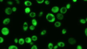
Reporter genes, such as green fluorescent protein (GFP), luciferase, or ß-galactosidase can be used to analyze transfection efficiency because their expression can be easily monitored. They can also be used to standardize transfection efficiencies between different transfection experiments by comparing the expression levels of their products. Reporter genes can be used alone or fused to a gene of interest to determine the protein expression level, the number of positive cells, or the location of the protein being studied.
Flow Cytometry

A flow cytometer can examine protein expression levels and transfection efficiency simultaneously. It captures data at the single cell level, enabling the rapid determination of the percentage of positive cells within a transfected cell population (% positive cells) (Nakatsuka et al. 2024). Additionally, expression levels on a per-cell basis can be determined by measuring the intensity of fluorescent signal associated with each cell.
Flow cytometry can detect the expression of a fluorescent reporter gene or directly detect the expression of a protein of interest, provided an antibody specific to that protein is available. With its extensive multiplexing capabilities, it can also be used for monitoring the cells expressing a protein of interest in mixed populations, such as blood samples, and for detecting multiple reporter genes or antibody-labeled proteins. Similarly, flow cytometry can be used to measure the biologic effect of modified cells in vitro and in vivo, for example, by monitoring the proportion and viability of B cells in PBMCs following incubation with B cell reactive CAR T cells. Additionally, a flow cytometer with sorting abilities can purify cells based on any measured characteristic, or group of characteristics, for example, allowing researchers to purify cells that express high levels of a transgene while discarding low or negative expressors.
Advantages
- Determines percentage of positive cells (quantitative measurement)
- Expression level on a per cell basis is given (quantitative measurement)
- Can enrich for positive cell population with sorting ability
- Can determine cell viability
- Facilitates high-throughput analysis
Limitations
- Relies on fluorescence, requiring either a fluorescent reporter gene or an antibody specific to the protein of interest conjugated to a fluorescent dye
- Requires the use of multiple controls for spectral compensation or unmixing and absolute quantification
- High initial cost of equipment and ongoing servicing
Western Blot Analysis

Western blot analysis can be used to identify or quantify the total expression of your transfected gene from a population of cells. With western blot analysis, you can determine the relative expression level of your transfected protein in a population of cells (Shalaby et al, 2022).
The process involves separating proteins by gel electrophoresis, transferring them onto a membrane, and detecting the target protein using specific antibodies. An internal control, such as GAPDH or ß-actin, is essential to ensure accurate and reliable quantification. Imaging systems that offer high sensitivity, accuracy, and reproducibility, enhance the quantitative analysis of gene delivery efficiency and provide clear evidence of total gene expression.
Advantages
- Inexpensive
- Gives confirmation of protein size and structure
Limitations
- Must be able to label and detect protein
- Time consuming due to running and transfer of gels and labelling protein for detection
- Cannot give expression information for individual cells
- For quantitative analysis, need an internal control and densitometer
Real-Time Quantitative PCR

Real-time quantitative PCR (qPCR) is a powerful tool for post-transfection analysis of cells. It enables precise quantification of transgene expression levels or the efficacy of gene silencing experiments (Schijeide et al. 2023).
This method is particularly advantageous because it requires minimal starting material, making it suitable for scenarios where transfected cells are available in limited quantities. By measuring the relative abundance of specific messenger RNA (mRNA) compared to a control, real-time qPCR provides insights into the gene expression changes induced by transfection. However, it is important to note that while real-time qPCR can quantify mRNA levels, it does not directly measure protein expression, which means any issues occurring post-mRNA synthesis, such as during protein translation, will not be detected.
Advantages
- High sensitivity
- Commonly used and widely accepted technique
Limitations
- Protein is not directly measured therefore issues occurring post-mRNA synthesis are not detected
- Moderately expensive
- Cannot give expression information for individual cells
Droplet Digital PCR

Droplet digital PCR (ddPCR) is an advanced technique used for post-transfection analysis of cells. It involves partitioning a sample into thousands of nanoliter-sized droplets, each acting as an individual PCR reaction. This method allows for the absolute quantification of target DNA or RNA molecules without the need for standard curves, providing high precision and sensitivity (Wu et al. 2024).
ddPCR is particularly useful for detecting low-abundance targets and offers greater tolerance to inhibitors compared to traditional qPCR. By measuring the number of positive droplets, researchers can accurately determine the expression levels of transfected genes, making ddPCR a valuable tool for assessing gene delivery efficiency and transfection success. Additionally, recent advances in ddPCR technology have facilitated greater multiplexing.
Advantages
- Provides absolute quantification of target DNA or RNA molecules
- High precision and sensitivity, especially for low-abundance targets
- Greater tolerance to inhibitors compared to traditional qPCR
- Does not require standard curves for quantification
Limitations
- Protein is not directly measured or identified, any issues occurring post-mRNA synthesis are not detected
- Cannot give expression information for individual cells
- More complex and time-consuming compared to traditional qPCR
Methods Summary
Table 1: Post-Transfection Cell Analysis Options
| Cell isolation | Quantification | Multiplexing | Cost | Single cell analysis | Throughput | Measures | |
|---|---|---|---|---|---|---|---|
| Cell counting | No | + | - | + | No | ++ | Viability |
| Flow cytometry | Yes | ++ | +++ | +++ | Yes | +++ | Protein and viability |
| Western blotting | No | ++ | + | + | No | + | Protein |
| Real-time PCR | No | ++ | ++ | ++ | No | ++ | RNA/DNA |
| ddPCR | No | +++ | +++ | ++ | No | + | RNA/DNA |
Featured Content
Transfection Resources
Introduction to Transfection
This section provides an overview of different transfection methods, transfection workflows, factors affecting transfection efficiency, and protocols.

Transfection Protocol Library
The Transfection Protocol Online Library contains protocols obtained from the literature, developed by Bio-Rad scientists, or submitted by scientists like you. Browse protocols to view our library and find your starting point.

An Optimized Workflow for Generating Anti-CD19 CAR T Cells by mRNA Electroporation
Here we describe the generation of anti-CD19 CAR-T cells using the GenePulser Xcell Electroporation System in an mRNA electroporation-based CAR-T workflow optimized for efficiency and viability.
Featured Content
References
Chong ZX et al. (2021). Transfection types, methods and strategies: a technical review. PeerJ 9, e11165.
Nakatsuka E et al. (2024). Characterization of DNA damage repair pathway utilization in high-grade serous ovarian cancers yields rational therapeutic approaches. Transl Oncol 50, 102119.
Gallicano GI et al. (2022). Molecular targeting of vulnerable RNA sequences in SARS CoV-2: identifying clinical feasibility. Gene Ther 29, 304–311.
Shalaby KE et al. (2022). Rapid assessment of CRISPR transfection efficiency and enrichment of CRISPR induced mutations using a dual-fluorescent stable reporter system. Front Genome Ed 4, 854866.
Schijeide BM et al. (2023). Determining on-target, off-target, and copy number status of transgenic events after CRISPR/Cas9 targeted AAVS1 safe-harbor modification of iPSCs using double-control quantitative copy number PCR. Curr Protoc 3, e635.
Wu X et al. (2024). Development and validation of a droplet digital PCR method for quantifying lentiviral vector infectious titer. Heliyon 10, e38512.
Featured Content
Related Content
Videos
- Using an Automated Cell Counter to Simplify Gene Expression Studies: siRNA Knockdown of IL-4 Dependent Gene Expression in Namalwa Cells
- Using the Gene Pulser MXcell Electroporation System to Transfect Primary Cells with High Efficiency
- Preparation of Gene Gun Bullets and Biolistic Transfection of Neurons in Slice Culture