
Western Blot Learning Center
Instruction for all steps in the western blotting workflow.
Western blotting is a powerful technique that allows you to positively detect your proteins, estimate quantities, and determine their molecular weights. All from a starting mixture of proteins extracted from cells or tissues.
The entire western blotting workflow consists of several individual steps, each of which is critical to producing high-quality data.
This Learning Center provides in-depth information on the theory and practice of each step of the western blotting process, starting from sample preparation to analysis of the final blot

-

Sample Preparation
Cells containing your protein of interest must be lysed completely to ensure a high yield while removing non-protein components of cells. Good sample preparation techniques ensure proteins remain undamaged for downstream analysis.
-
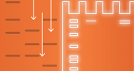
Electrophoresis
Electrophoresis separates the proteins in the sample and provides molecular weight data for detected proteins during subsequent detection.
-

Transfer
Separated proteins are transferred from the gel to a membrane where they are immobilized. Efficient protein transfer is required for maximum western blot sensitivity.
-

Detection
A good, clean western relies on the specificity and sensitivity of your antibodies. In addition to detection of specific proteins, the total protein in a sample can also be visualized with total protein stains or newer technologies that eliminate the need for staining and destaining.
-

Image Acquisition
Accurate imaging of your western blot is crucial for capturing blot data for downstream analysis. Understanding basic imaging concepts such as sensitivity, resolution, and sources of background noise can help you maximize image data quality.
-
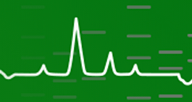
Image Analysis and Quantification
An image of a western blot is rich with information. Some experiments require only a qualitative answer, but properly designed and executed western blot experiments can also provide quantitative data on relative protein expression between samples.
Receive Western Blotting Tips and Online Resource Updates
Sign Up to Be the First to Know
Receive Western Blotting Tips and Online Resource Updates
Keep up to date with useful tips to continuously improve your western blotting experiments from sample preparation through image analysis. Sign up to be the first to be notified when new western blotting resources like tips and tricks, posters, protocols, webinars, and how-to-videos become available.
-
Image

Blot Talk: Monthly Live Webinar Series.
Join our live Blot Talk webinars to troubleshoot real western blots submitted by you, discover the latest techniques and advancements, and learn essential best practices. Get your questions answered live and gain practical advice to get the best results from your western blots every session!
-
Image
Stain-Free Western Blotting Guide
Find out how Stain-Free technology can revolutionize your western blotting.
-
Image
Western Blot Selector Tool
Find the right products for you using the free Western Blot Selector Tool.
-
Image
Protein Standard Temporary Tattoo
Fun and free protein standard tattoo.
-
Image
Electrophoresis & Western Blotting Layout Post-It
This simple tool allows users to keep track of their Western Blotting experiment from sample preparation to imaging.
-
Image
Stain-Free Western Blotting Resource Collection
Learn how you can master Stain-Free Western Blotting using this comprehensive literature resource collection.


