TC20 Automated Cell Counter

TC20 Automated Cell Counter

The TC20 Automated Cell Counter can quickly and accurately count cells in suspension. It can be used with or without trypan blue for viable cell or total cell counts on a broad range of cell types and sizes. Counting can be performed significantly faster than manual methods whilst improving reproducibility.

Read More
Ordering Information

Contact a Specialist

Overview

The TC20 automated cell counter counts cells in one simple step using its innovative auto-focus technology and sophisticated cell counting algorithm to produce accurate cell counts in less than 30 seconds. Upon insertion of a counting slide, the TC20 cell counter rapidly provides a total cell count (with or without trypan blue staining) and assesses cell viability via trypan blue exclusion.

Key Features and Benefits

  • Compatible with a broad range of cell sizes and types — counts cell lines, primary cells, and stem cells
  • Innovative auto-focus technology — removes the variation associated with manual focusing and leads to precise cell counts in 30 seconds
  • Flexible cell size gating — user selects a population of interest in complex samples, such as primary cells, or lets the cell counting algorithm do all the work
  • Accurate cell viability analysis — analyzes cells using multifocal plane analysis
  • Easy to archive and analyze — stores up to 100 counts in the onboard memory for access any time, or use the optional TC20 data analyzer software on your PC to further analyze exported cell images

Getting all the data you need about your cell cultures is fast and easy; the TC20 cell counter and disposable counting slides eliminate the need for tedious setup, cleaning, or maintenance. The TC20 cell counter is simple and intuitive to use — learn to use the TC20 automated cell counter in the introduction video or take a virtual test drive.

  • TC2 load

    Load

    Load the sample onto a slide.

  • TC20 insert

    Insert

    Insert the slide into the TC20 cell counter; counting automatically begins.

  • TC20 View

    View results

    Obtain a total cell count (without trypan blue) or total and live cell counts (with trypan blue) in 30 seconds.

Get Accurate and Reproducible Results

The TC20 automated cell counter uses microscopy with auto-focus that analyzes multiple focal planes to identify the best plane. Without requiring any user input, the sophisticated cell counting algorithm uses the image acquired from the best focal plane to identify cells and exclude debris, thereby calculating the total cell count. The auto-focus leads to highly reproducible cell counts with reduced user-to-user variability compared to a hemocytometer and cell counters with manual focus. Using auto-focus instead of subjective manual focusing is especially important when assessing cell viability because an incorrectly selected focal plane will lead to inaccurate results.

Unlike cell counters based on electrical impedance (Coulter principle), the TC20 image-based analysis allows the user to view the cells in real time, providing visual proof that the TC20 cell counter has correctly identified cells.

The TC20 automated cell counter provides accuracy comparable to results obtained with a hemocytometer. It can count cells with a 6–50 μm cell diameter and within a broad concentration range of 5 x 10⁴–1 x 10⁷ cells/ml, which eliminates the need to dilute cells, thus reducing errors associated with sample dilutions prior to counting. Read more about how the TC20 compares to a hemocytometer.

This automated counter uses disposable counting slides with a patent-pending design that ensures even distribution of cells throughout the counting chamber, regardless of the user's pipetting style, allowing every user to accurately and consistently count cells.

For complex samples composed of multiple cell populations, such as primary cells, users can adjust the cell size gates to define the population of interest that will be counted. When counting multiple sample replicates, the TC20 automated cell counter can save the position of the cell size gates and apply them to subsequent counts.

Size Gating Image

When user-defined gates are enabled you can select a population of interest by adjusting the position of the cell size gates.

Cell Viability

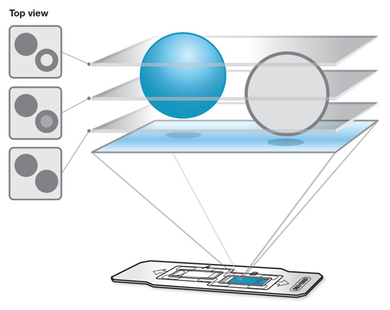
The TC20 automated cell counter uses multifocal plane analysis to assess cell viability. The conventional method of analyzing viability using a single focal plane can lead to inaccurate conclusions because light scattering and the alignment of cells at different heights in a counting chamber can change the appearance of cells — live cells may appear to be dead and vice versa. To determine whether cells are viable, the TC20 counter analyzes each cell using images acquired from multiple focal planes during the focusing step. Find out more about the advantages of multifocal plane analysis.

Helpful Viewing

TC20 Automated Cell Counter System Tour

A quick overview of the main features of the TC20 automated cell counter. Eliminate the tedium of manual cell counting, see how the TC20 counter provides a total count of mammalian cells and a live/dead ratio in one simple step, giving accurate, reproducible results in less than 30 seconds.

Introducing the TC20 Automated Cell Counter

Using the TC20 Automated Cell Counter

See how The TC20 Automated Cell Counter makes cell counting simple with automatic multiple focal plane analysis and trypan blue detection, yielding improved accuracy and reducing the undercounting of live cells. See how size gates can be manually adjusted, and data can be exported for further analysis on a standalone PC.

Counting Cells with Bio-Rad's TC20 Automated Cell Counter

TC20 Gallery

TC20 Cell Gallery: images of compatible cell types including cell lines, primary and stem cells

Minimize Your Sample Preparation

The broad concentration range of the TC20 automated cell counter eliminates the need to dilute cells prior to counting, reducing the errors associated with sample dilutions that may be necessary when counting cells by other methods. The counting algorithm discriminates and counts individual cells within clusters of up to five cells, providing accurate counts without the need to extensively declump cells prior to loading.

Product Details

The TC20 automated cell counter has two counting modes. Depending on the sample type, the user-defined gates can be either enabled or disabled.

Samples with Multiple Cell Populations (stem cells and other primary cells)

User-defined gates must be enabled. Once the user-defined gates are enabled, a histogram will be displayed at the beginning of each count and the user can select the population of interest by adjusting the position of the cell size gates. Only objects within the designated range will be analyzed as cells; those with diameters outside of the size range will be excluded from the cell count.

If the user-defined gates are disabled, the cell counting algorithm will automatically determine the population to count without any user input. The algorithm will count the cells of the most predominate cell type that typically comprise the largest peak, which may not be the population the user wants to count.

  • Setup Window Image
    Gating setup window.
  • Size Gating Image
    Cell size gating window.

To make counting of such samples easier, when counting multiple sample replicates, the TC20 cell counter can save the positions of gates and apply them to subsequent counts (choose Yes in the “Use saved gates” field in Gating setup).

If the Image preview is On, an image of cells will be displayed during gating. In the image, only cells with diameters between the selected size gates will be marked with a yellow dot.

Immortal Cell Lines

When counting immortal cell lines or other samples that are composed of cells with similar cell size, the user-defined gates can be disabled. The cell counting algorithm then automatically identifies the cell population of interest without requiring user input.

The TC20 automated cell counter uses microscopy with auto-focus that analyzes multiple focal planes to identify the best plane. The cell counting algorithm then uses the image acquired from the best focal plane to identify cells and exclude debris, thereby calculating the total cell count.

Using auto-focus instead of subjective manual focusing is especially important when assessing cell viability because an incorrectly selected focal plane will lead to inaccurate results.

If trypan blue is detected along with the total cell count, the TC20 counter assesses cell viability. The conventional method of analyzing viability using a single focal plane can lead to inaccurate conclusions because light scattering and the alignment of cells at different heights in a counting chamber can change the appearance of cells — live cells may appear to be dead and vice versa. To determine if cells are viable, the TC20 counter analyzes each cell using images acquired from multiple focal planes during the focusing step.


Multifocal Plane Image


Effect of multifocal plane analysis on live/dead count accuracy for HeLa cells.

Viability Assessment Live Cells, % Dead Cells, %
Single best focal plane* 35 65
Multiple focal planes* 65 35

* On single focal plane, 35% of live cells were misidentified as dead cells.


View, Export, and Analyze Your Data

Cell Display Image

After viewing cell count results, users can choose to view an image of the counted cells on the TC20 automated cell counter with the ability to zoom in on an image with the click of a button.

 
Data Export Image

After a count, a JPEG file of the cell image is available for immediate export to a USB flash drive or, to save every cell image, automatic data export can be enabled. Each cell image contains a metatag with the count data and can be further analyzed using TC20 data analyzer software under Download Tab. This optional software can be used to review single images, perform multi-file comparative analyses, and generate reports.

 
Previous Counts Image

Results from 100 counts are stored in the TC20 cell counter so you can always go back and recover counts based on their acquisition date and time stamp. Previous count results can be exported via the USB port and opened in a Microsoft Excel spreadsheet, allowing easy comparison of results between experiments.

 
Current Count Image

You can assign a custom sample name that will be associated with the count result and the JPEG image. To save time when working with multiple sample replicates, enable the automatic name serialization option to populate numerically serialized names.

 
TC20 with printer image

The optional thermal label printer can be connected to the TC20 counter to print results onto labels that can be placed into a laboratory notebook for traceability of the count results.

 
Dilution Calculator Image

Users can access the onboard dilution calculator to determine volume adjustments required to achieve the cell concentration needed for the next experiment.


The data below show the TC20 automated cell counter is compatible with a broad range of cell types, such as cell lines, primary cells (from tissue or blood), and stem cells. The TC20 cell counter can count cells with a 6–50 µm cell diameter and within a broad concentration range of 5 x 10⁴–1 x 10⁷ cells/ml, which eliminates the need to dilute cells, thus reducing the error associated with sample dilutions prior to counting.

Cell Type Graph Image
The TC20 automated cell counter demonstrates accurate cell counts across a range of cell sizes. Small (PBMC, Jurkat), medium (HeLa), and large (MEF) cells were counted with a hemocytometer, a TC20 automated cell counter, and a competitor's image-based automated cell counter. The TC20 counter and hemocytometer cell counts showed no statistically significant differences. Precision is indicated by the standard deviations; error bars represent average standard deviations. Cell counts on the TC20 counter were performed on one instrument with four sample replicates.
 
Cell Viability Graph Image
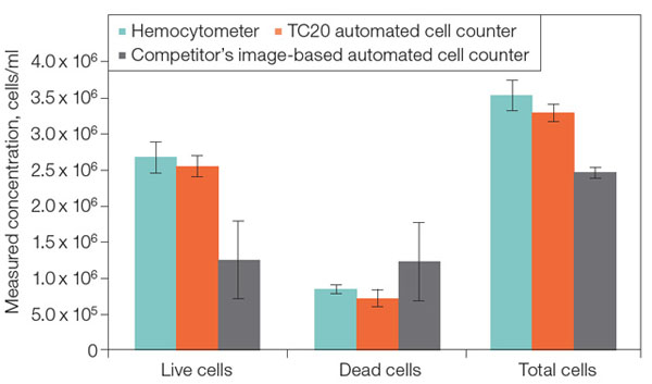
The TC20 automated cell counter demonstrates accurate counts of viable cells. Pan T cells mixed with trypan blue (1:1) were counted with a hemocytometer, a TC20 automated cell counter, and a competitor’s image-based automated cell counter. The TC20 counter and hemocytometer cell counts showed no statistically significant differences. Precision is indicated by the standard deviations; error bars represent average standard deviations. Cell counts on the TC20 counter were performed on four different instruments with five sample replicates.

Further Reading

Mammalian tissue or cell culture techniques were developed in the early 1900s as a method of maintaining animal organs outside of the body. During the 1940s and 1950s cell culture technology emerged as an essential tool to support virology research by enabling vaccine production through viral purification. Today, cell culture is used worldwide as a model for a variety of studies, including intra- and intercellular communication, tissue/organ development or organization, and different disease states, such as cancer, Parkinson's disease, and diabetes.

  • Cell Biology Cell Biology 101

    In this guide the most common cell types used in cell biology are described.

  • Cell Culture Basic Cell Culture

    This presentation provides an introduction to growing and maintaining mammalian cells in culture


Specifications

Counting time, sec
30
Cell concentration range, cells/ml
5 x 10⁴–1 x 10⁷
Optimal cell concentration range, cells/ml
1 x 10⁵–5 x 10⁶
Cell diameter range, µm
6–50
Sample volume, µl
10
Data storage, counts
100
Data export
Via USB flash drive
Printer connectivity
Yes
Dilution calculator
Yes
Image of cells available
Yes
Dimensions (W x D x H), cm (in)
19 x 15 x 25.4 (7.5 x 6 x 10)
Weight without the external power supply, kg (lb)
2.2 (4.8)

Use

For Research Use Only. Not for use in diagnostic procedures.

Ordering

items

Use the filters below to refine results!

Image
cell counting feature
1450102

120–240 V, includes instrument, power supply, USB flash drive, 30 dual-chamber counting slides (60 counts), 1.5 ml trypan blue

Image
145-0103
1450103

120–240 V, includes instrument, power supply, USB cable, USB flash drive, thermal label printer, 1 roll of 185 labels, 30 dual-chamber counting slides (60 counts), 1.5 ml trypan blue

Accessories

Image
145-0003_tn.jpg
1450003

30 slide pack of dual-chamber slides (60 counts), includes 1.5 ml trypan blue, for use with TC10™ or TC20™ automated cell counter

Image
145-0015_tn.jpg
1450011

30 slide pack of dual-chamber slides (60 counts), each slide can provide counts for 2 separate samples or dilutions, for use with TC10™ or TC20™ automated cell counter

Image
145-0015_tn.jpg
1450015

5 x 30 slide pack of dual-chamber slides (300 counts), each slide can provide counts for 2 separate samples or dilutions, for use with TC10™ or TC20™ automated cell counter

Image
145-0015_tn.jpg
1450016

10 x 30 slide pack of dual-chamber slides (600 counts), each slide can provide counts for 2 separate samples or dilutions, for use with TC10™ or TC20™ automated cell counter

Image
145-0015_tn.jpg
1450017

20 x 30 slide pack of dual-chamber slides (1,200 counts), each slide can provide counts for 2 separate samples or dilutions, for use with TC10™ or TC20™ automated cell counter

Image
145-0015_tn.jpg
1450018

30 x 30 slide pack of dual-chamber slides (1,800 counts), each slide can provide counts for 2 separate samples or dilutions, for use with TC10™ or TC20™ automated cell counter

Image
145-0015_tn.jpg
1450019

40 x 30 slide pack of dual-chamber slides (2,400 counts), each slide can provide counts for 2 separate samples or dilutions, for use with TC10™ or TC20™ automated cell counter

Image
145-0015_tn.jpg
1450020

80 x 30 slide pack of dual-chamber slides (4,800 counts), each slide can provide counts for 2 separate samples or dilutions, for use with TC10™ or TC20™ automated cell counter

Image
145-0021_tn.jpg
(Discontinued) Trypan Blue
1450021
5 x 1.5 ml, sufficient for 750 counts (10 µl/count), 0.4% in 0.81% sodium chloride and 0.06% potassium phosphate dibasic solution, sterile filtered, for use with TC10™ or TC20™ automated cell counter
Image
145-0022_tn.jpg
(Discontinued) Trypan Blue
1450022
10 x 1.5 ml, sufficient for 1,500 counts (10 µl/count), 0.4% in 0.81% sodium chloride and 0.06% potassium phosphate dibasic solution, sterile filtered, for use with TC10™ or TC20™ automated cell counter
Image
145-0014_tn.jpg
(Discontinued) System Test Kit
1450014

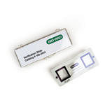
Kit for validation of TC10™ or TC20™ automated cell counter functionality, includes verification slide, instructions

Image
145-0005_tn.jpg
1450005

120–240 V, connects to the TC10™ or TC20™ automated cell counter, for printing results onto labels, includes thermal label printer, USB cable, 1 roll of 500 labels

Image
145-0007_tn.jpg
1450007

Pkg of 1 roll, 500 labels, for use with thermal label printer for printing results obtained with the TC10™ or TC20™ automated cell counter

Downloads

TC20™ Data Analyzer Software
SOFT-TC20-Data-Analyzer
TC20 data analyzer software, compatible with JPEG image files from the TC20 automated cell counter
TC20™ Automated Cell Counter Firmware
SOFT-TC20-FW

Firmware update for the TC20 cell counter, version 2.103

Documents

Number Description Options
10024423
Instruction Manual, TC20 Automated Cell Counter
10024424
Quick Guide, TC20 Automated Cell Counter
6283
TC20 Automated Cell Counter Brochure
[ Add to Cart (Free) ]
6282
TC20 Automated Cell Counter Product Information Sheet, Rev A
10018213
TC20 and TC10 Validation Kit Instruction Guide
6003
Comparison of Count Reproducibility, Accuracy, and Time to Results between a Hemocytometer and the TC20 Automated Cell Counter
6087
Comparison between the TC10 Automated Cell Counter and the Lactate Dehydrogenase (LDH) Assay to Assess Cellular Toxicity in Vitro, Rev A
6152
Accurate Counting of Bio-Plex Magnetic or Polystyrene Beads Using the TC10 Automated Cell Counter, Rev A
6777
Protocol for Single Cell Gene Expression Profiling by Multiplex RT-qPCR
6340
A Comparison of the TC10™ Automated Cell Counter and the Hemocytometer for Quantifying Insect Hemocytes, Rev A
6853
Reliable Single-Cell Sorting with Bio-Rad's S3e™ Cell Sorter Application Note