
Introducing the Western Blot Learning Center
Perfect your western blotting. Learn from the experts.
On This Page | Normalization Methods | Stain-Free Imaging | New Publication Guidelines | Stain-Free Western Workflow | ChemiDoc Imaging System | Resources | Related Products |
Western Blot Normalization Methods
A quantitative western blot accurately measures changes in protein expression by making relative comparisons between different treatments.
A properly designed quantitative western blotting experiment ensures that:
- The amount of protein loaded is proportional to the signal intensity
- All quantified targets' signal intensities fall within the linear range of the imaging system
- Common blot-to-blot variations are accurately assessed by an internal loading control
Western blot normalization allows you to faithfully compare changes in protein expression by establishing the baseline needed to correct against common errors such as inconsistent sample preparation, pipetting error, and uneven protein transfer. Proper western blot normalization is required to show that the changes in band intensities correlate to the biological changes in your samples.
Conventional loading controls rely on consistent levels of a reference protein in each sample. Typically, a "housekeeping protein" (HKP) such as β-actin, β-tubulin, or GAPDH is used as a loading control, with the assumption that the expression levels of these proteins remains constant.
The Problem with Housekeeping Proteins
Reliable assessment of the changes in target protein expression levels requires the measurement of both the target protein and the loading control protein in their linear dynamic ranges for immunodetection. Unfortunately, housekeeping proteins are usually highly expressed, whereas target proteins are often expressed only in low abundance. Thus, to detect the target protein of interest, large amounts of cell lysate may need to be loaded, resulting in overloading of HKPs, yielding oversaturated reference bands, out of their linear range. Furthermore, HKP expression levels may not be constant but instead vary with different experimental treatments and other factors.
Variation in Housekeeping Protein (HKP) Expression Levels
The expression level of housekeeping proteins can change due to:
- Experimental conditions
- Developmental changes
- Post-transcriptional regulation
- Differences in cell
- Tissue age & type
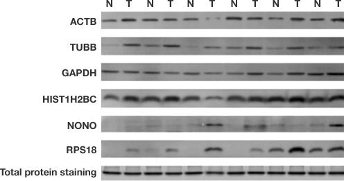
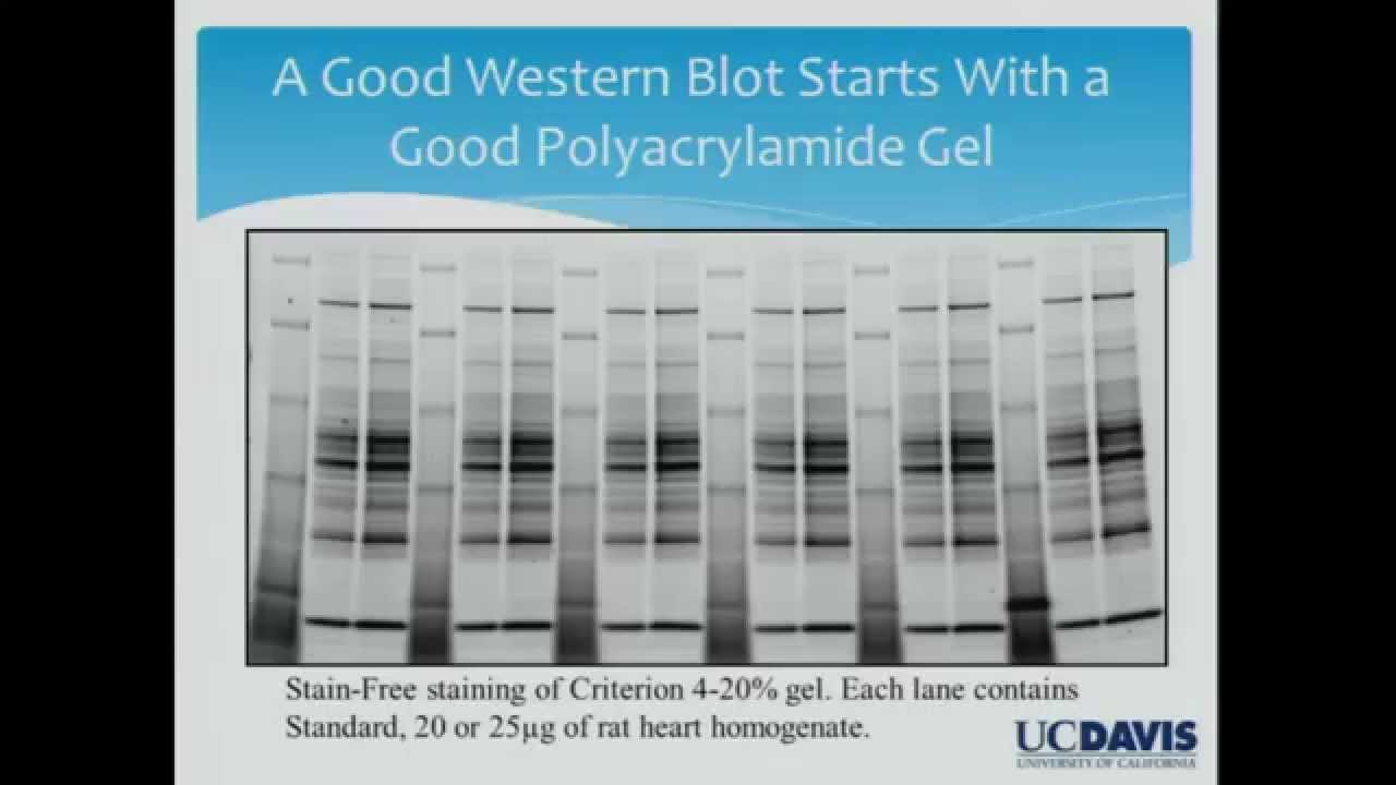
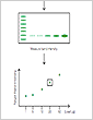
Differences in five candidate housekeeping proteins and total protein staining between tumor and non-cancerous tissues in the validation sample set. Immunodetection measurements of housekeeping protein levels show poor linearity and do not accurately indicate cell lysate loading levels. Total protein normalization of HKPs yields consistent band intensities. Hu X et al. (2016). Common housekeeping proteins are upregulated in colorectal adenocarcinoma and hepatocellular carcinoma, making the total protein a better "housekeeper". Oncotarget 7, 66, 679–66, 688.
Total Protein Normalization: The Better Alternative
The use of total protein measurement for western blot loading controls (total protein normalization; TPN) is a method devised to resolve the inherent difficulties with linearity in the immunodetection of both target and control proteins. Total protein levels can be determined by staining the membrane with total protein stains, e.g., Coomassie, SYPRO Ruby, Flamingo, amido black, or Ponceau S.
Because total protein stains are less sensitive than antibody-based immunodetection, they are far less likely to result in an oversaturated signal. As a result, they exhibit good linearity in the common loading range of 10–50 μg of cell lysate. Therefore, both low-abundance target protein levels, measured using sensitive immunodetection techniques, and high-abundance housekeeping protein levels, measured using less sensitive total protein staining techniques, can each be measured in their respective linear dynamic ranges.
Unfortunately, the use of total protein stains can interfere with subsequent blot development and visualization steps, typically requiring tedious staining and destaining procedures, a major reason for the lack of widespread adoption of total protein normalization using these stains. The invention of stain-free imaging technology has made total protein normalization a much better alternative for quantitative western blotting.
Advantages of Total Protein Normalization
Stain-free imaging allows for total protein normalization.
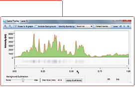
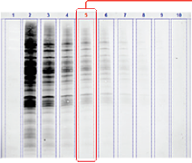
Lane profiling of total protein signal helps remove background and refine band detection.
Total protein normalization takes into account:
- The intensity of all proteins in the lane
- Sample loading variations
- Variations during electrophoresis
- Variations during transfer
Further Reading: Total Protein Normalization


Stain-Free Imaging
Stain-free imaging technology utilizes a proprietary trihalo compound to enhance natural protein fluorescence by covalently binding to tryptophan residues with a brief UV activation. Images of the gel or membrane after transfer can easily be captured multiple times without staining and destaining steps. This allows visualization, verification, and validation at all steps of electrophoresis and blotting, potentially saving time wasted on western blots with problems that would not otherwise have been detected until the later stages of blot processing and development.
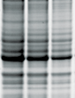
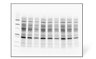
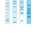
Verification of Western Blotting Protein Transfer Using Stain-Free Imaging
Gel before transfer
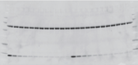
Membrane after transfer
Gel after transfer
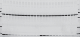
Assessment of protein transfer using a stain-free enabled imaging system. Images of the gel before and after transfer and of the membrane after transfer were taken using the Criterion Stain-Free imager. Serial 1:2 dilutions of hemoglobin (starting quantity, 80 ng), with 1.8 μg of BSA/lane as a carrier (top band), were electrophoretically separated on a 4–20% 26-well Criterion Stain-Free gel.
Using stain-free total protein measurement as the loading control, researchers can ensure that both the target protein and loading control are measured in the linear dynamic range in a typical western blot experiment. Furthermore, stain-free imaging allows for the complete elimination of the inherently problematic use of housekeeping proteins as loading controls on western blots. Stain-free total protein measurement serves as a more reliable loading control than housekeeping proteins, particularly in the loading range commonly used for cell lysates, permitting the user to obtain truly quantitative western blot data by normalizing bands to total protein in each lane.
Learn More About Stain-Free Technology »
A Better Way to Normalize Quantitative Western Blots
Total protein normalization using stain-free imaging technology allows normalization across a wider dynamic range.

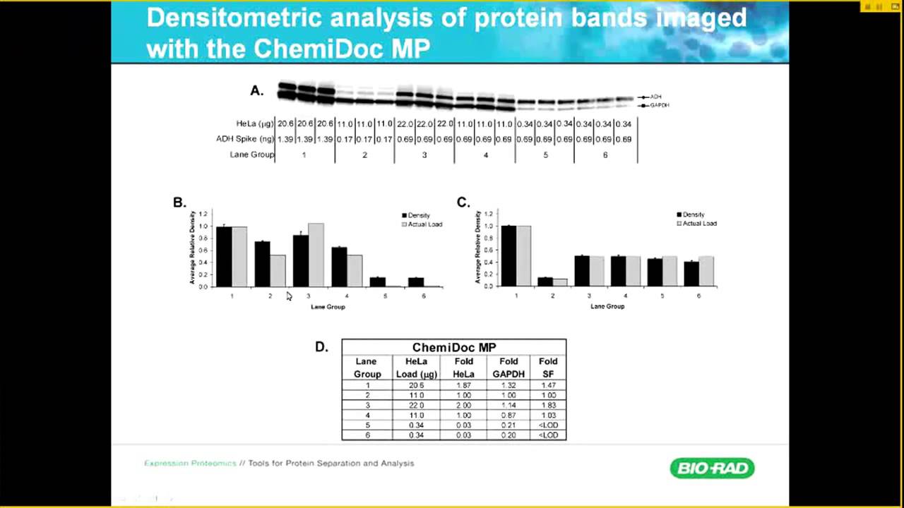
Linearity comparison of stain-free total protein measurement and immunodetection of three housekeeping proteins in 10–50 μg of HeLa cell lysate. On the left are representative images of (a), stain-free blot and the chemi blots for (b), β-actin; (c), β-tubulin and (d), GAPDH. Lane labels correspond to total protein load (μg). Although the actin and tubulin signals appear linear, the densitometric ratio was far below the predicted “quantitative response” of actual loading whereas the stain-free signal correlated to the expected result (e). Taylor SC Posch A (2014). The design of a quantitative western blot experiment. Biomed Res Int. 36, 1590
Further Reading: Stain-Free Imaging for Western Blotting

Trends in Protein Separation and Analysis: The Advance of Stain-Free Technology
This article focuses on the theory and concepts surrounding stain-free technology as well as other traditional methods of protein staining.
New Publication Guidelines
Several major scientific journal publishers have recently revised their editorial guidelines for data publication, with an emphasis on reproducibility and quantitation criteria. The Journal of Biological Chemistry guidelines for collecting and presenting data specifically address quantitative western blot publication, providing guidance around normalization methods and supporting data, citing concerns with the use of housekeeping proteins, imaging technology, and antibody specificity.
Bio-Rad's Stain-Free Western Workflow meets the new publication guidelines – does yours?
Meeting the New Publication Requirements for Quantitative Western Blotting
| JBC Guidelines: Quantitative Blots | How to Meet Requirements | Bio-Rad Western Workflow Solutions |
|---|---|---|
| "Housekeeping proteins should not be used for normalization without evidence that experimental manipulations do not affect their expression" | Use Total Protein Normalization (TPN) instead of housekeeping proteins | The Stain-Free Western Workflow suite simplifies stain-free TPN. This technology is proven with over 100 publications, allows for improved accuracy and will make your data more reliable |
| "Methods including detection of enhanced chemiluminescence using X-ray film have a very limited dynamic range" | Use an imaging system with at least 4 logs of dynamic range | ChemiDoc Systems are stain-free imagers with the sensitivity of film Image Lab Software enables simple image generation and quantitation |
| "A description of the data supporting the specificity of all antibodies is required" | Use fully validated antibodies | PrecisionAb Antibodies offer documented specificity and validation protocols |
Further Reading: Publication Guidelines
- Nature: Information for Authors & Referees > Policies > Image Integrity and Standards
- Fosang AJ, Colbran RJ (2015). Transparency Is the Key to Quality. J Biol Chem, 290(50): 29, 692–29, 694
- McNutt M (2014). Journals Unite for Reproducibility. Science 346(6210): 679
- Yadav G, Oh K (2018). Defining the New Normal in Quantitative Western Blot Data. Bioradiations.com
Key Factors for Publishing High-Confidence Western Blot Data
Prof. Aldrin V. Gomes (University of California, Davis) relies heavily on biophysical proteomic techniques in his studies of signal transduction, including western blotting. As an editor of several journals, he has routinely observed the submission of low-quality western blot data, leading him and other scientists to question the accuracy of protein quantification by western blotting in prior publications.
Prof. Gomes has deeply examined key success factors in western blotting, including antibody selection and reducing errors and misinterpretations by implementing a standardized procedure and proper controls. Here he presents key factors for high-confidence western blot data:
- Antibody specificity and blocking reagent quality
- Properly selected and executed controls
- Total protein loading control vs. housekeeping protein loading control
Prof. Gomes also presents tips to reduce the overall bench time required for first-rate western blots and how to minimize the pain of going through a long procedure only to obtain equivocal results. As the old saying goes, If you didn't have time to do it right the first time, when will you find time to do it again?
Stain-Free Western Blotting
Stain-free western blotting allows you to quickly check electrophoresis and blot transfer quality and obtain truly quantitative western blotting results, updating traditional blotting techniques with innovative tools.
Faster Results. Better Data.
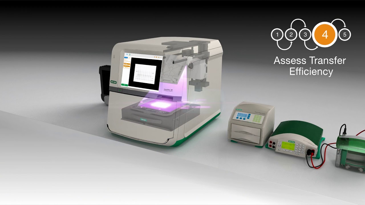
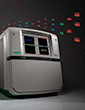
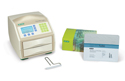
ChemiDoc Imaging System
Imagine No Compromises
With the ChemiDoc MP Imaging System, the superior quantitation of digital imaging meets the sensitivity of film. This best-in-class system images and analyzes gels and western blots. It is designed for multiplex fluorescent western blotting, chemiluminescence detection, general gel documentation, and stain-free technology imaging needs.
Better Options. Better Results.
Stain-Free
Total Protein Normalization
Learn how to properly design a quantitative western blotting experiment and faithfully compare protein expression levels, without concern for changing or overexpressed HKP’s.
See Data »
Chemiluminescence
Replaces Film
Save money on consumables and reduce environmental waste without compromising on performance. Get the sensitivity of film and the convenience of digital documentation.
See Data »
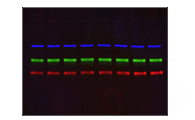
Multiplex Fluorescence
3 Colors
Save sample, avoid the errors commonly associated with stripping, reprobing, and cutting blots and answer more complex biological questions by optimizing your experiments to fluorescence.
See Data »
Resources
FEATURED WEBINARUnderstand the Factors That are Crucial to Successful Western Blotting

Join Bio-Rad western blotting expert Kenneth Oh and his guest speakers for a webinar series that explores the many factors that go into the design and execution of successful and repeatable western blots. Chemiluminescence or fluorescence, qualitative or quantitative blot, not only will we discuss the how-to's, we'll take a deeper dive and discuss the why's.
View Webinar
Reliable Quantification of Western Blot Data
Dr. S.C Taylor compares results generated for identical blots with camera-based imaging technologies and film. Learn common pitfalls associated with film and densitometric analysis.
Designing a Quantitative Western Blot Experiment to Avoid Common Pitfalls
The quantitative western blot methodology is described with a focus on solid experimental design and data analysis to achieve reproducible, publication quality results in the shortest time frame.

Quantitative Western Blotting: Optimization is the Key
Each step of the quantitative western blot workflow is discussed with a focus on optimization to assure quantitative, reproducible final data that reflect the experimental conditions.

Western Blot Selector Tool
Find the right products for you using the free Western Blot Selector Tool.

Western Blot University
Courses designed to make you a western blotting expert.
Image Lab
Image Lab Touch Software Resources
Bio-Rad provides instructional videos for Image Lab Touch Software 2.4, covering the primary applications of the ChemiDoc MP Imaging System. Topics include, logging in, Smart Tray Technology, capturing your image, and exporting data.
Image Lab Software Resources
Bio-Rad provides tutorials for basic acquisition and advanced analysis use of Image Lab Software 6.0.1. Topics include, densitometric analysis, molecular weight, normalization, purity assessment, and more!
Application Guides

Western Blot Doctor Troubleshooting Guide
Our self-help troubleshooting guide covers solutions to many common and not-so-common western blotting issues and helps your blots look their best.

Protein Blotting Guide
(PDF 7.9 MB)Details on blotting technology, available products, and methods, plus tips, techniques, and troubleshooting.

Electrophoresis Guide
(PDF 6.4 MB)A guide to polyacrylamide gel electrophoresis and protein detection, including theory, product selection, protocols, and more.
Documents Library
ChemiDoc MP System Brochure
(PDF 1.26 MB)Explore the features and benefits of the new high-end imaging system for the best fluorescence and chemiluminescence detection.
Stain-Free Approach to Western Blotting
(PDF 864 KB)Learn how to use stain-free technology for total protein normalization as an alternative to the standard blot normalization process.
Stain-Free Western Blotting Brochure
(PDF 593 KB)See how the Stain-Free Western Workflow compares to traditional western blotting workflows.
Determining the Appropriate Sample Load When Performing a Stain-Free Western Blot
(182 KB)
Validating the Expression Consistency of a Housekeeping Protein
(198 KB)
Publications
A Defined Methodology for Reliable Quantification of Western Blot Data
Learn about the methodology to obtain reliable quantitative data from chemiluminescent western blots using standardization procedures coupled with the updated reagents and detection methods.
The Design of a Quantitative Western Blot Experiment
Read a summary about a complete western blot workflow with a focus on sample preparation and data analysis for quantitative western blotting.
Related Products
Fluorescent Western Blotting Products
Bio-Rad's fluorescent western blotting workflow is a seamless integration of products designed to work together to offer guaranteed results.
Western Blotting
Western blotting products include the Stain-Free Western Workflow, protein transfer systems, membranes, filter paper, blotting buffers, and detection kits
Mini-PROTEAN TGX Stain-free Precast Gels
Mini-PROTEAN TGX Stain-Free Precast Gels for polyacrylamide gel electrophoresis (PAGE) allow for fast run times, high transfer efficiency, and rapid detection of proteins with stain-free enabled imagers.
PrecisionAb Validated Western Blotting Antibodies
These premium antibodies are lab-validated using strict testing criteria to ensure superior performance in western blotting detection.
Trans-Blot Turbo Transfer System
The Trans-Blot Turbo System is a rapid protein transfer apparatus that can transfer protein to membrane in as little as 3 minutes. Trans-Blot Turbo Transfer Packs provide greater transfer efficiency in less time.
Protein Ladders and Standards (Markers)
Prestained and unstained molecular weight standards for protein electrophoresis applications including SDS-PAGE, western blotting, 2-D PAGE, and isoelectric focusing (IEF).

Clarity ECL
Clarity family of Western ECL Substrates provides simple, high-performance solutions for all your western chemiluminescence needs.
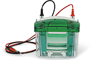
Electrophoresis Chambers
Our electrophoresis chambers enable rapid, high-resolution protein separation on precast or handcast gels over a variety of different gel sizes.
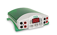
Power Supplies
From basic to high-voltage capacity, our power supplies provide simple programming in a compact, stackable case.