
On This Page |
See What You’ve Been Missing | Sensitivity | Putting It All Together | Resources | Related Products |
Do You Know How Image Quality Affects Data Quality?
Western blotting experiments have always been challenging, requiring the optimization of multiple procedures to obtain good results. Western blots are currently evolving from a qualitative test to a quantitative assay, demanding practitioner expertise in blotting, detection, and imaging techniques as well as data validation.
Just as experiments require optimization, building quality imaging systems requires expertise. Over the 35+ years Bio-Rad has been developing quality imaging systems, we’ve learned a lot. As researchers have moved from film to digital imaging and expanded detection methods to chemiluminescence and fluorescence, our R&D team has continually improved the materials we use, explored design parameters, and optimized the processes behind our advanced development, all with one goal: to build the most sensitive western blot detection system in the world.

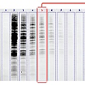
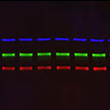
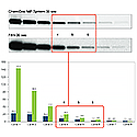
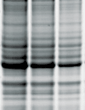
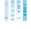
Good vs. Poor Instrument Design. Images of the same blot taken using the IR channel on two different imaging systems. A. image captured on competitor instrument; B. image captured on a ChemiDoc MP System.
Carefully optimized instrument design prevents image artifacts and high background. A low-cost imaging system may save money initially, but wasted time and poor results are costly, too. Poor image quality affects your ability to see and quantitate your data. Ultimately, your data quality is what matters most. All parts of the ChemiDoc MP System have been selected for maximum signal, low noise, and ultimately the best data quality. These component materials, mechanicals, camera design, lens, filters, illumination, and software have been optimized to work together and give you the most accurate blot imaging system available.
See What You've Been Missing
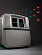
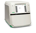
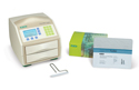
The New ChemiDoc MP Imaging System:
Under the Hood
Learn how Bio-Rad imaging systems are custom- designed for peak performance from the leaders of our Imaging R&D team. See what differentiates a merely good imager from a truly great imaging system.
Limitations in an imaging system may not be readily apparent to the user. While obvious imaging artifacts are easily noticed, faint or missing bands, poor sensitivity, inconsistent results, inaccurate quantitation, and other issues may be mistaken as simply the way an instrument functions. Researchers focused on their biological questions shouldn't also need to be experts in imaging. The end user generally assumes that an instrument just works the way it’s supposed to.
But that can be a risky assumption, and doing good science means accounting for all sources of error. Imaging is an experiment too, and needs experimental controls in place for accuracy. For example, will you get the same results when a blot is placed on different areas of the platen?
Our R&D team has done as much of that work for you as possible, by examining all possible sources of error then refining and optimizing the components, optics, electronics, and software to yield consistent results.
Sensitivity
Signal to Noise: What's in Your Background?
The definition of sensitivity in image quantification is measurable signal above background noise. In western blot imaging, it's the ability to see a very faint band clearly above any background. To accomplish this, the system must clearly identify what is relevant signal. This proposition sounds simple but is actually rather complicated. In a digital imaging system, what the camera detects as signal is not necessarily what should be quantified in an assay, but the camera doesn’t natively know the difference between signal and noise, it just collects light.
Both signal and noise arise from multiple sources, making it difficult to differentiate and correct for them. To ensure what you are quantifying is only the signal from your assay — and that the signal is quantified correctly — we get a little fanatical about some pretty unusual things. We've tried to discover every source of error, analyze its effects, and mitigate them all.
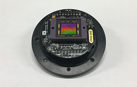
Refined camera electronics The Bio-Rad-designed camera powering the ChemiDoc MP System has exceptionally low read noise and dark current in order to minimize background and maximize sensitivity.
When you are imaging a gel or blot, the sensor inside of the camera converts light to electrical signals. The sensor will read any source of light, regardless of whether it is from the sample or from light leaking into the instrument. A light leak occurs when the camera or imaging system has a gap that allows light to penetrate into the normally light-tight chamber, exposing the sensor to extra light.
Bio-Rad has been committed to designing light-tight imaging systems since the launch of Gel Doc (2000) and ChemiDoc (2001) Imagers, continuing with significant developments over many years of investment through the imaging portfolio. The new ChemiDoc MP is our most light-tight system yet.
The Right Light
Filtering is challenging in a camera-based imaging system. Light comes in from all angles, and the image sensor must be protected from stray light. Understanding the complexities of light paths and balancing the principles of glass optics is not straightforward, requiring trial and error for continuous improvement. Our first-generation ChemiDoc MP System had only one filter in front of the lens. The new instrument actually has four different levels of filtering: a filter behind the lens, two filters in front of the lens, and the lens itself acts as a spatial filter that removes all the highly off-angle light and parasitic light. The more sophisticated filtering in the new ChemiDoc MP Systemis really what enabled us to improve performance, especially in the near-infrared. This opens up possibilities for NIR applications that previously would have required a separate system.
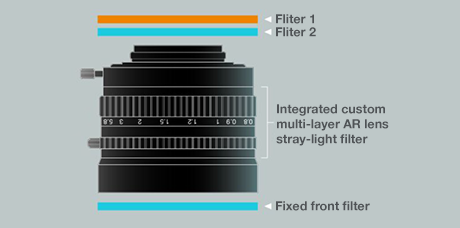
Custom lens and filter stack The new lens features three external filters and specialized internal coatings for off-angle light suppression.
No existing lens met our stringent optical requirements, so we had a custom lens developed, which yielded resolution higher than what was available from off-the-shelf lenses.
We also optimized the anti-reflection coatings and added specialty coatings not found in stock lenses, including special black materials on the lens housing interior that help mitigate parasitic stray light that can bounce around within lenses. The final result is significantly reduced background levels.
Autofluorescence: What's Glowing?
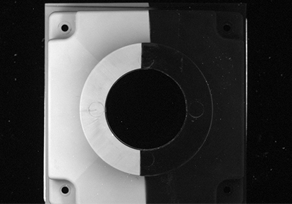
Autofluorescenece Suppression: Solarization due to UV exposure over
time caused the uncoated part of this component to fluoresce, which
would have caused increased background in the images.
Many materials will fluoresce when exposed to high intensity light, termed autofluorescence. With fluorescent western blots, the blot and some of the interior of the instrument is subjected to intense excitation light. If any materials show autofluorescence and emit light within the bandpass of the emission filters, then background levels of the image will increase. This results in lower overall sensitivity of the system.
For the ChemiDoc MP System, we selected materials and coatings for the interior surfaces specifically to reduce fluorescence and thus eliminate this source of background.
This new coating also provides resistance to solarization, in which a material exhibits changes in color or emissivity after exposure to ultraviolet light, but can also progressively degrade the physical or mechanical properties of many materials, especially polymers, after prolonged exposure.
Putting It All Together
The New ChemiDoc MP Imaging System
The careful design and proprietary software algorithms in the ChemiDoc MP System yield uniform data for every application, in every position. Together with our patented image-correction algorithms, the custom lens, filter strategy, uniform LED illumination, and interior coatings make a huge difference in imaging performance versus competitor systems.
The ChemiDoc MP System was designed to meet the needs of scientists. With all detection options available and a great user interface, you can choose whichever method works best for you experiments, and pursue answers where your research leads you, without being limited by your imaging technology.
Imagine No Compromises
With the ChemiDoc MP Imaging System, the superior quantitation of digital imaging meets the sensitivity of film. This best-in-class system images and analyzes gels and western blots. It is designed for multiplex fluorescent western blotting, chemiluminescence detection, general gel documentation, and stain-free technology imaging needs.
Resources
Introducing the Western Blot Learning Center
Perfect your western blotting. Learn from the experts.

Featured WebinarUnderstand the Factors that are Crucial to Successful Western Blotting
Join Bio-Rad western blotting expert Kenneth Oh and his guest speakers for a webinar series that explores the many factors that go into the design and execution of successful and repeatable western blots. Chemiluminescence or fluorescence, qualitative or quantitative western blot, not only will we discuss the how-tos, we'll take a deeper dive and discuss the whys.
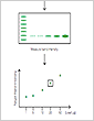
Total Protein Normalization
Learn how total protein normalization is key to obtaining quantitative western blot data for publication.

Stain-Free Imaging Technology
Find out how stain-free imaging can improve your protein electrophoresis and western blotting protocols.
Fluorescent Western Blotting
Fluorescence western blotting and total protein normalization can facilitate quantitative and accurate protein expression analyses.
Film vs. Digital Western Blot Imaging
Modern digital imaging systems with wide dynamic range enbale accurate quantitation over a range of signal intensities equal to or better than film.
Image Lab Touch Software Resources
Bio-Rad provides instructional videos for Image Lab Touch Software 2.4, covering the primary applications of the ChemiDoc MP Imaging System. Topics include, logging in, Smart Tray Technology, capturing your image, and exporting data.
Image Lab Software Resources
Bio-Rad provides tutorials for basic acquisition and advanced analysis use of Image Lab Software 6.0.1. Topics include, densitometric analysis, molecular weight, normalization, purity assessment, and more!
Application Guides

Western Blot Doctor Troubleshooting Guide
Our self-help troubleshooting guide covers solutions to many common and not-so-common western blotting issues and helps your blots look their best.

Protein Blotting Guide
(PDF 7.9 MB)
Details on blotting technology, available products, and methods, plus tips, techniques, and troubleshooting.
Electrophoresis Guide
(PDF 6.4 MB)
A guide to polyacrylamide gel electrophoresis and protein detection, including theory, product selection, protocols, and more.
Documents Library
ChemiDoc MP System Brochure
(PDF 1.26 MB)Explore the features and benefits of the new high-end imaging system for the best fluorescence and chemiluminescence detection.
Stain-Free Approach to Western Blotting
(PDF 864 KB)Learn how to use stain-free technology for total protein normalization as an alternative to the standard blot normalization process.
Stain-Free Western Workflow Brochure
(PDF 593 KB)See how Stain-Free western blotting compares to traditional western blotting workflows.
Determining the Appropriate Sample Load When Performing a Stain-Free Western Blot
(182 KB)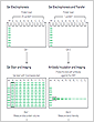
Validating the Expression Consistency of a Housekeeping Protein
(198 KB)
Publications
-
A Defined Methodology for Reliable Quantification of Western Blot Data
Learn about the methodology to obtain reliable quantitative data from chemiluminescent western blots using standardization procedures coupled with the updated reagents and detection methods.
-
The Design of a Quantitative Western Blot Experiment
Read a summary about a complete western blot workflow with a focus on sample preparation and data analysis for quantitative western blotting.
Related Products
ChemiDoc Imaging Systems
ChemiDoc Imagers offer best-in-class performance for fluorescence and chemiluminescence detection and all general gel documentation applications.

Image Lab Software
Image acquisition and analysis software for Bio-Rad Gel Doc, ChemiDoc, and GS-900 Systems. Capture and analyze digital image data from electrophoresis gels and blots.
Western Blotting
Western blotting products include protein transfer systems, membranes, filter paper, blotting buffers, and detection kits.
Fluorescent Western Blotting
Bio-Rad's fluorescent western blotting workflow is a seamless integration of products designed to work together to offer guaranteed results.
Trans-Blot Turbo Transfer System
The Trans-Blot Turbo System is a rapid protein transfer apparatus that can transfer protein to membrane in as little as 3 minutes. Trans-Blot Turbo Transfer Packs provide greater transfer efficiency in less time.
PrecisionAb Validated Western Blotting Antibodies
These premium antibodies are lab-validated using strict testing criteria to ensure superior performance in western blotting detection.
Mini-PROTEAN TGX Stain-Free Precast Gels
Mini-PROTEAN TGX Stain-Free Precast Gels for polyacrylamide gel electrophoresis (PAGE) allow for fast run times, high transfer efficiency, and rapid detection of proteins with stain-free enabled imagers.
Protein Ladders and Standards (Markers)
Prestained and unstained molecular weight standards for protein electrophoresis applications including SDS-PAGE, western blotting, 2-D PAGE, and isoelectric focusing (IEF).

Clarity ECL Substrates
Clarity family of Western ECL Substrates provides simple, high-performance solutions for all your western chemiluminescence needs.
SureBeads Protein G Magnetic Beads
SureBeads Protein G Magnetic Beads enable fast, easy, consistent immunoprecipitation without centrifugation.