

Powerful Protein Detection Without Lysate Preparation
See How In-Cell Western Assays Can Speed Up Your Research
Introduction to In-Cell Western Assays
In-Cell Western (ICW) is a powerful quantitative and qualitative technique that enables the analysis of protein expression in-situ without the hassle of cell lysate sample preparation.
-
The In-Cell Western User:
In-Cell Western is ideal for those who have already validated the specificity of their antibodies with traditional western blotting or for those interested in overall protein expression without needing to account for molecular weight.
-
Uses for In-Cell Western:
- Quantification of protein expression changes in response to drug treatments or genetic manipulations
- Evaluation of protein-protein interactions
- Screening large libraries of compounds in drug discovery and development
- Analysis of protein localization within specific cellular compartments
Example of a Typical In-Cell Western Workflow

Templates & Protocol
Download these pre-made plate templates and optimized protocol to simplify your In-Cell Western Image Lab Analysis:
Protocol Length Comparison: In-Cell Western vs Traditional Western Blot
The In-Cell Western Assay is an efficient, time-saving method ideal for those who have already validated the specificity of their antibodies with traditional western blotting or for those interested in overall protein expression without needing to account for molecular weight.
-

Total Time for In-Cell Western (up to 384 samples)

-

Total Time for Traditional Western Blot (up to 24 samples)

Advantages of In-Cell Western Assay vs. Traditional Western Blotting

Advantage 1:
Shorter protocol — no need for time-consuming sample preparation, electrophoresis, and transfer steps.

Advantage 2:
Quantification of multiple targets (up to 3-plex) in numerous samples (up to 384-wells).

Advantage 3:
Protein detection in situ — qualitative and quantitative immunofluorescence.
In-Cell Western Assays Made Easy Using the ChemiDoc MP and ChemiDoc Go Imaging Systems
The top reasons why the ChemiDoc Imaging System family is superior to competitor imagers for In-Cell Western imaging:
- Advanced Multiplexing: Detect up to 2 targets with the ChemiDoc™ Go Imaging System or up to 3 with the ChemiDoc™ MP Imaging System — surpassing competitor systems that typically max out at 2 targets.
- Broad Dye Compatibility: Unlike systems limited to NIR dyes, the ChemiDoc MP Imaging System supports full-spectrum imaging, while the ChemiDoc Go Imaging System is fully compatible with StarBright™ Dyes.
- Exceptional Image Quality: Delivers high-resolution images and integrates seamlessly with Image Lab™ Software for streamlined data analysis.
- Ready-to-Use Controls: When paired with our hFAB™ Rhodamine Housekeeping Protein Fluorescent.


Simplify your workflow with EveryBlot™ Blocking Buffer — the all-in-one blocking buffer that’s now compatible with Western Blotting, ELISA, and In-Cell Western™.
- Low background, high signal: Optimized to minimize nonspecific binding, leading to cleaner images and more accurate quantification — crucial for ICW, where signal clarity is vital.
- Multiplex compatibility: Compatible with fluorescent detection and multiplexing, making it ideal for use with the ChemiDoc Imaging Systems, especially when detecting multiple targets simultaneously.
- Broad antibody compatibility: Designed to work well with a wide range of primary and secondary antibodies, including both fluorescently labeled and traditional antibodies.
- Phosphoprotein detection: Contains no phosphate-based buffers, making it ideal for use with phospho-specific antibodies.
- Consistent performance across cell types: Validated for use in various adherent and suspension cell lines, enhancing reproducibility across experiments.

Take your In-Cell Western™ assays to the next level with StarBright™ Blue 700
- Unmatched sensitivity — powered by novel polymer-based chemistry, StarBright™ 700 delivers signal intensity comparable to femtogram-level ECL detection.
- Exceptional signal-to-noise ratio — achieve clearer results and cleaner backgrounds, even with low-abundance targets.
- Optimized for multiplexing — seamlessly integrate StarBright™ 700 into your dual- or triple-target detection workflows with ChemiDoc™ Go or ChemiDoc MP Imaging Systems.

Find the Right hFAB Rhodamine Housekeeping Protein Fluorescent Primary Antibody for Your Experiment
Bio-Rad offers commonly used house-keeping primary antibodies conjugated to a rhodamine dye, making it easy to add to any assay as a natural control.
- Simple — no requirement for a secondary antibody; one-step detection of housekeeping proteins using highly cross-adsorbed antibodies for high specificity and sensitivity
- Versatile — compatible with use of a wide range of other fluorophores including StarBright Blue 700 and DyLight 800 Fluorophores for multiplex western blotting
- Unique — HuCAL Technology eliminates cross-reactivity with primary/secondary antibodies from other species
- Diverse — antibodies have been tested to detect human or mouse housekeeping proteins
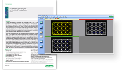
Image Lab Analysis of In-Cell Western Assays
Image Lab Software simplifies In-Cell Western analysis by taking advantage of its harmonious pairing with any ChemiDoc Imager, making it a powerful yet easy-to-use package for acquisition and analysis of your In-Cell Western Plate. By using the downloadable plate templates provided on this page or creating your own using the Volume Tool, Image Lab Software automatically calculates volume intensity of each well. When coupled with Bio-Rad’s hFAB Rhodamine Housekeeping Protein Fluorescent Primary Antibodies, you can set control wells along with blank wells that are used for background subtraction to calculate adjusted volume intensity. In-Cell Western analysis allows for quantification of multiple targets using spectrally distinct fluorescent dyes and is a quick method for finding relative protein levels in numerous samples.
Applications & Technologies
Protocol & Templates
Download these pre-made plate templates and optimized protocol to simplify your In-Cell Western Image Lab Analysis:
-
Image

Western Blotting
Learn about the powerful technique that allows you to positively detect your proteins, estimate quantities, and determine their molecular weights.
-
Image
Western Blot Learning Center
The Western Blot Learning Center is a complete reference on all of the steps of western blotting, includes practical theory, protocols, and recommendations on how to make your blots better from experts.


Western Blot University
Courses designed to make you a western blotting expert.

