The understanding, manipulation, and use of human adult stem cells as therapeutic agents is an important and growing field of research. Unlike embryonic stem cells, research with adult stem cells does not involve the creation, use, or destruction of human embryos and is therefore far less controversial. The therapeutic benefits of adult stem cell transplantation are well established. None more so than in the treatment of leukemia, where they are used to reconstitute the immune system following immune ablation (Crees et al. 2023). Whilst tissue replacement therapies continue to show promise, adult stem cells have also been posited as important sources of regenerative and immunosuppressive mediators (Leclerc et al. 2022, Tolstova et al. 2023).
Types of adult stem cells and the tissue they form include:
- Hematopoietic stem cells (HSCs) — blood
- Mesenchymal stem cells (MSCs) — bone, cartilage, adipose tissue
- Neural stem cells (NSCs) — neurons and glia
- Limbal epithelial stem cells — corneal epithelium
Adult stem cells are rare. They exist in relatively small numbers in their respective niches making the study of these cells in vivo challenging. However, flow cytometry and cell sorting are useful tools for their identification and characterization because they allow analysis at the single cell level based on the expression of cell surface markers that can be detected following labelling with specific antibodies conjugated to fluorophores. Hematopoietic stem cells (HSCs) and mesenchymal stem cells (MSCs) are both particularly amenable to study using flow cytometry.
Already know what you're looking for?
Discuss with a flow cytometry specialist.
Hematopoietic Stem Cells
In adults, HSCs are located primarily in the bone marrow but canalso circulate in the blood under certain conditions. They give rise to the vast majority of cells found in the blood, including all leukocytes (white blood cells) and erythrocytes (red blood cells). In human bone marrow HSCs are present at less than 1% of the total cellularity.
Identifying HSCs Using Flow Cytometry
There are several markers that can be used to identify HSCs, many of which are species dependent. In humans, the phenotype lineage marker negative (Lin-)/ CD34+/CD38-CD45RA-/CD90+/CD49f+ is the most widely accepted strategy for identification of HSCs. However, the expression of CD34+/CD90+ is sufficient to identify the cell population exclusively responsible for multilineage reconstitution following immune ablation. Further simplification can be achieved by only considering CD34+ cells and thus including progenitor cells, which, whilst not fulfilling the definition of stem cells, nevertheless contribute to immune reconstitution.
Table 1. Human HSC and progenitor markers.
| Positive markers | Negative markers | |
|---|---|---|
| HSCs | CD34, CD90, CD49f, Sca-1 | Lin, CD38, CD45RA, |
| Multipotent Progenitors | CD34, Sca-1 | Lin, CD38, CD45RA, CD90, CD49f |
Mesenchymal Stem Cells
Also known as mesenchymal stromal cells and multipotent stromal cells, debate continues as to the most appropriate label for MSCs. However, there is consensus that MSCs are able to differentiate into several cell types in vitro. These include osteoblasts, which are responsible for forming bone, chondrocytes, which make up cartilage, and adipocytes, which are responsible for the storage of fat. It is widely accepted that MSCs are present in the bone marrow as well as several other tissues, including dental pulp, adipose tissue, and the umbilical cord. In human bone marrow, MSCs are typically present at less than 0.02% of the total cellularity.

Identifying MSCs Using Flow Cytometry
The most widely accepted definition of MSCs are cells that are tissue culture plastic adherent and express CD73, CD90, and CD105 but lack expression of CD11b, CD14, CD19, CD34, CD45, CD79a, and HLA-DR. They must also have the potential for differentiation into cells able to form bone, cartilage, and adipose tissue. However, the above phenotypic definition based on cell surface marker expression is insufficient to specifically identify MSCs in vivo. Indeed, no single marker or collection of markers has been shown to exclusively identify all MSCs in vivo, and tissue localization strongly influences marker expression. In human bone marrow, sorting using CD271 and/or CD73 has proved highly selective for MSCs.
Table 2. Human tissue-specific MSC markers.
| All tissues | Markers |
|---|---|
| Culture expanded positive markers | CD73, CD90, CD105 |
| Culture expanded negative markers | CD11b, CD14, CD19, CD34, CD45 CD79a, HLA-DR |
| Bone marrow positive markers | CD271, CD73, SSEA-4, Stro-1, CD146, GD2 |
| Adipose tissue positive markers | CD271, CD34, CD146, GD2 |
| Umbilical cord positive markers | CD146, CD49f, GD2 |
Challenges of Stem Cell Analysis Using Flow Cytometry
The relative rarity of stem cells in vivo is a persistent challenge hampering their study. Although flow cytometers can typically analyze thousands of cells per second, this relative rarity can lead to excessively long acquisition times. The speed at which data can be collected on any system is dependent on the number of cells that can be passed through the interrogation point and successfully electronically processed in a given time.
Reagent choice is also an important consideration. In general, when analyzing rare cells, it is prudent to use a fluorophore that is bright enough to easily stand out above the increased noise associated with larger data files. This issue is exacerbated in the case of MSCs because analysis often involves enzymatic digestion of solid tissues, a process that can generate substantial autofluorescent debris. The exceptional brightness of StarBright™ Dyes makes them ideal for this application.
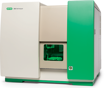
Using the ZE5 Cell Analyzer for Stem Cell Research
The speed at which data can be collected using the ZE5 Cell Analyzer is the key advantage that sets it apart from other cytometers in the field of stem cell research. There are a number of factors that make this possible.

- Industry leading event rate — the ZE5 Cell Analyzer can collect 100,000 events per second without electronic aborts, whereas most instruments start to struggle at just 20,000. This allows a significant number of cells to be collected quickly even when working with a very rare cell type.
- Clog resistant fluidics — the ZE5 Cell Analyzer has a reversible sample pump and automatic clog detection. A high detection rate is dependent on fluidics that don’t clog easily and lead to instrument down time, with the ZE5 Cell Analyzer you can be more confident when working at high event rates.
- On board temperature control and sample vortexing — during long acquisition runs it is essential to keep fresh samples cool and vortexing is required prior to starting acquisition of each new sample. The ZE5 Cell Analyzer allows both these steps to be automated giving you the confidence to walk away.
ZE5 Cell Analyzer Stem Cell Publications
References:
- Crees ZD et al. (2023). Motixafortide and G-CSF to mobilize hematopoietic stem cells for autologous transplantation in multiple myeloma: a randomized phase 3 trial. Nat Med 29, 869-879.
- Leclerc K et al. (2022). Hox genes are crucial regulators of periosteal stem cell identity. Development 150, dev201391.
- Tolstova T et al. (2023). The effect of TLR3 priming conditions on MSC immunosuppressive properties. Stem Cell Res Ther 14, 344.


