Western Blot Doctor™ — Protein Band Appearance Problems

The Western Blot Doctor is a self-help guide that enables you to troubleshoot your western blotting problems. In this section, you can find solutions to issues related to protein band appearance.

Other sections in the Western Blot Doctor:

Problems and Solutions


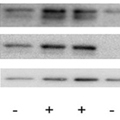
Problem: Inconsistent control protein levels among samples

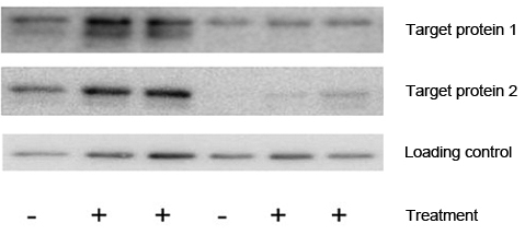
Variation observed among the loading controls in each lane

Western blot with varying levels of control proteins – Western Blotting Doctor - Loading Control Issues
Possible causes: Solutions:

Samples may have different amounts of total protein

  • Check that total protein levels are consistent:
  • To determine whether the changes in loading control levels are due to differences in the amount of sample loaded, or if the differences are caused by variations in expression of the loading control proteins, use total protein stains (e.g., Ponceau S, Coomassie, colloidal gold, or SYPRO Ruby) to visualize proteins on gels and blots before and after transfer to determine relative protein loading. If planning to use the blot in downstream steps, make sure that your stain can be removed or is compatible with antibody detection.  For example, Coomassie and colloidal gold are not compatible with downstream steps (see Bio-Rad Protein Stains and the Protein Stain Selection Guide)
    • To determine if there is residual, untransferred protein remaining on the gel, use a total protein stain on the gel after transfer
    • To verify protein transfer, stain the membrane with Ponceau S after blotting
    • Visualize total protein on gels and blots using Bio-Rad’s Stain-Free Gels featuring our proprietary Stain-Free Technology. In addition to providing visual verification of transfer at every step, stain-free imaging enables total protein normalization in each lane, eliminating the need for housekeeping proteins as loading controls

Loading control protein levels may vary between test and control conditions

  • Check that loading control expression is consistent across conditions using a secondary loading control. If loading control expression varies with experimental conditions, try using another loading control
  • Try total protein normalization using stain-free technology instead of normalizing to a single housekeeping protein. For more information see the following:
 

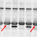
Problem: Ghost protein bands

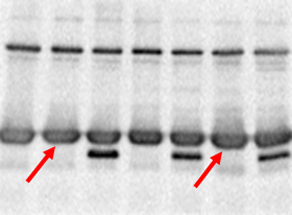
Nonspecific protein bands, can be large or out of place.

A blot with ghost bands – Western Blot Doctor - Signal Saturation Issues
Possible causes: Solutions:
Sample overloading
  • Decrease total protein loaded for samples
  • Check concentration of protein samples (e.g., using Bradford or Lowry protein assays) before loading the gel
  • Optimize sample loading; see Determining the Appropriate Sample Load for Western Blots Protocol

Too much antibody

  • Decrease concentration of primary and/or secondary antibodies
  • Optimize your primary and/or secondary antibody concentrations using a checkerboard screening protocol

Exposure too long

  • Use a shorter exposure time
  • Use multi-acquisition feature on data acquisition software

Rapid substrate consumption

  • Use a less sensitive detection substrate
  • Reduce incubation time with detection substrate

Blot was moved

  • When constructing the blotting sandwich, do not readjust the blot after the gel has come in contact with the membrane, as this can lead to ghosting on the blotting membrane.
 

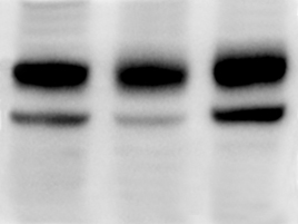
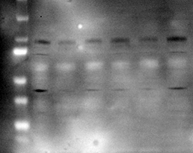
Problem: Swirls or missing bands; bands appear diffuse on blot

Bands do not look flat, may be trailing off in multiple directions.  Bands may look broad and fuzzy.

Possible causes: Solutions:

Contact between the membrane and the gel was poor; air bubbles or excess buffer remain between the blot and the gel (see also Blot Background > White spot)

  • Carefully move the roller over the membrane in both directions until air buffers or excess buffer are removed from between gel and membrane and complete contact is established
  • Use thicker filter paper in the gel/membrane sandwich
    (see our Blot Absorbent Filter Paper selection)
  • Replace the foam pads. Pads compress and degrade with time and will not hold the membrane to the gel
    Foam pads for Bio-Rad wet tank blotting systems:
 

Problem: Blurry bands

(see also Protein Band Size and Pattern > Band(s) at slightly higher MW than expected)

Possible causes: Solutions:

Gel run at too high a voltage

  • Repeat gel electrophoresis at lower voltage
  • Run at lower voltage for entire run
  • Run at lower voltage until proteins begin to enter the resolving gel, then increase voltage for remainder of run
PowerPac™ Power Supplies can be programmed to automatically run methods with up to nine steps

Trapped air bubbles present during transfer

  • Carefully remove air bubbles between the gel and membrane before protein transfer

Incorrect running buffer composition

  • Prepare fresh running buffer or use premixed commercial buffers (see our selection of Buffers and Reagents)
 

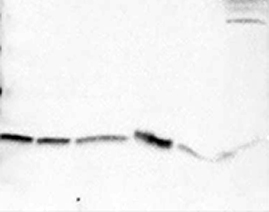
Problem: Bands are curved (smiling) not straight

 
Possible causes: Solutions:

Running conditions were too fast, gel became overheated

  • Check and optimize gel electrophoresis conditions. Consult your instruction manual or the Electrophoresis Guide
  • Reduce voltage during electrophoresis
  • Run gel at 4°C. Place electrophoresis cell in a 4°C cooler during run. Use chilled buffers, a cooling coil, or a “blue ice” cooling insert
 

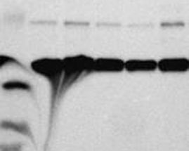
Problem: Broad or misshapen bands

 
Possible causes: Solutions:

Gel electrophoresis problems

  • Electrophoresis artifacts may occur as a result of poor gel polymerization, inappropriate running conditions, contaminated buffers, sample overload, etc. Consult your instruction manual for more details, and see the Electrophoresis Guide for guidance in all aspects of protein electrophoresis
  • Check the salt concentrations of the samples, especially when running salt-precipitated samples. When possible, maintain similar salt contents in all wells.  Wells with higher salt levels tend to expand when next to wells with less salt due to osmosis. High salt differentials (especially between sample and buffers) can also cause larger band distortion. Be careful when running salt-precipitated samples
  • High-salt samples can often be desalted using Bio-Gel® P6 columns
 

Problem: White (negative) bands on film using ECL method


Possible causes: Solutions:

Too much protein loaded

  • Load less sample
  • Repeat with dilution series of sample
  • Optimize the sample loading; see Determining the Appropriate Sample Load for Western Blots Protocol

Antibody concentration is too high

  • Reduce/optimize the antibody concentrations using checkerboard screening protocols
 

Problem: No bands

(see also Signal Strength Problems > Faint bands, weak or no signal) A blot with weak-signal – Western Blot Doctor - Weak-Signal Detection
Possible causes: Solutions:

Possible over-transfer or under-transfer
(see also Protein transfer or binding issues)

  • Confirm protein transfer by staining the membrane with Ponceau S and/or the gel with Coomassie Blue Dye, or use Bio-Rad’s Stain-Free Gels to verify transfer using our unique stain-free imaging technology
  • Note how well any prestained molecular weight markers have transferred onto the blot
  • Optimize and check transfer conditions and setup (especially orientation to electrodes)
  • Repeat using two membranes in case protein has transferred through the first membrane (over-transfer is especially likely with low-MW proteins)

Blocking agent interfering with signal

Buffers may contain sodium azide, which inactivates HRP

  • Use azide-free buffers

ECL detection reagents may be contaminated

  • Use fresh detection reagents

Peroxide may be inactive, resulting in lower peroxidase signal

  • Add fresh peroxide to substrate buffer

Inappropriate secondary antibody used

  • Retrace steps to check compatibility between primary and secondary antibodies
  • Reprobe with correct secondary or strip blot and reprobe if necessary
  • Repeat experiment with the correct antibody combination

Wrong concentration of antibody or low affinity to the target protein

  • Increase the antibody concentration 2–4 times higher than initial trial
  • Use a checkerboard screening protocol
  • Increase length of incubation
  • Reduce stringency of wash steps
  • Lower temperature, reduce detergent concentration, reduce ionic strength
  • Test and optimize antibody on dot blots
  • Try an alternate antibody. Bio-Rad now offers high-quality antibodies for all applications

Antibody not suitable for western blotting

  • Check datasheet for recommended conditions
  • Test and optimize antibody on dot blots
  • Validate target and antibody combinations using checkerboard screening protocols
  • Try an alternate antibody
Bio-Rad now offers PrecisionAb™ Validated Western Blotting Antibodies for superior performance in western blot detection.

Antibody activity loss due to long-term or improper storage

  • Test on a dot blot at several concentrations
  • Freeze aliquots of antibody and only thaw one at a time as needed for blots; store thawed aliquots at 4°C
  • Use fresh aliquots of antibody that have been stored at –20ºC or below
  • If storing an antibody for a very long period of time, store at –80ºC
  • Avoid repeated freeze-thaw cycles
  • For more information, see Tips for Caring for Your Antibody

Antigen not expressed in source material

  • Use another source of target protein
  • Include a positive control in experiment (all PrecisionAb Antibodies include a positive control lysate)

Not enough antigen loaded onto

 
Page Contents