
The Western Blot Doctor is a self-help guide that enables you to troubleshoot your western blotting problems. In this section, you can find solutions to issues related to protein band size and pattern problems.
Other sections in the Western Blot Doctor:
- Band Appearance Problems
- Blot Background Problems
- Signal Strength Problems
- Band Size and Pattern Problems
Problems and Solutions
Click on the thumbnail that is most representative of your own blot to discover the probable causes and find specific solutions to the problem.
Problem: Band(s) at lower MW than expected
| Possible sources of unexpectedly low-MW bands include protein cleavage or degradation, splice variants, and nontarget proteins bearing similar epitopes. | 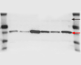 |
| Possible causes: | Solutions: |
| Target protein has been cleaved or digested |
|
| Splicing variant exists |
Bio-Rad now offers high-quality antibodies for all applications |
| Another protein bearing the same/similar epitope is detected by the antibody |
Bio-Rad now offers PrecisionAb™ Validated Western Blotting Antibodies for superior performance in western blot detection |
Problem: Band(s) at slightly higher MW than expected or blurry
| Bands at MW slightly higher than expected and/or blurred may indicate protein modifications such as glycosylation. | 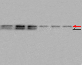 |
| Possible causes: | Solutions: |
| Protein may be glycosylated or otherwise modified at one or more amino acid residues |
|
Problem: Band(s) at significantly higher MW than expected
| Sources of unexpectedly high-MW bands include protein-protein interactions and antibody cross-reactivity. | 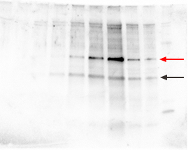 |
| Possible causes: | Solutions: |
| Dimers, multimers, or protein-protein interactions occurring because samples have not been fully reduced or denatured. |
|
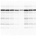
Problem: Multiple bands at various MWs
| Multiple nonspecific bands on the blot may be due to antibodies of poor quality or at too high a concentration, insufficient blocking, or nonspecific binding due to the presence of SDS. | 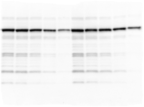 |
| Possible causes: | Solutions: |
| Primary antibody concentration too high or cross-reactivity with similar epitopes on other proteins |
|
| Secondary antibody concentration too high, leading to nonspecific binding |
|
| Protein exists in several different isoforms |
|
Primary or secondary antibody contaminated with nonspecific IgG or with IgG cross-reactive among species
|
|
| Monoclonal antibodies reacted nonspecifically with SDS-denatured proteins |
|
| Nonspecific interactions occurring due to ionic associations; for example, avidin, a glycosylated protein, may bind to more acidic proteins on blots |
|
| Insufficient blocking of nonspecific sites |
|
| SDS caused nonspecific antibody binding to immobilized proteins |
|