

PureBlu™ DAPI Nuclear Staining Dye #1351303
Pkg of 5 vials, 50 µg each, DAPI nuclear staining dye powder
Safety Data Sheet
Overview
Use PureBlu DAPI Dye for routine nuclear staining in fluorescence microscopy and cell imaging applications. PureBlu DAPI Dye is a highly pure formulation of DAPI in a user-friendly format allowing easy preparation of the working solution, with no weighing step and only a single dilution after resuspension.
DAPI (4',6-diamidino-2-phenylindole) is a well-characterized blue-emitting fluorescent compound widely utilized for nuclear staining. DAPI permeates cell membranes and binds to the minor groove of A/T-rich dsDNA sequences, thus preferentially staining nuclei (Figure 1).
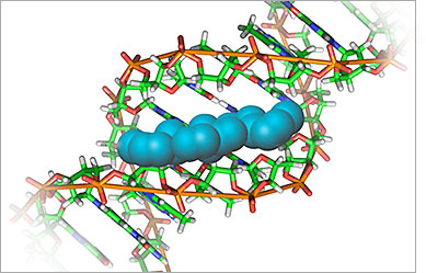
Fig. 1. DAPI binding dsDNA.
When bound to dsDNA, PureBlu DAPI Dye has a maximum excitation wavelength in the ultraviolet range (359 nm), and the dye can be optimally detected in the blue channel with an emission maximum of 461 nm (Figure 2). The characteristic Stokes shift between excitation and emission wavelengths is fairly wide for DAPI, making this dye an optimal choice when good spectral separation is desired to reduce fluorescence interference, for example, in chromatin counterstaining for immunofluorescence microscopy.
Fig. 2. DAPI excitation and emission spectra.
Related Products
More Information
Specifications
After resuspension: stable for 1 year at –20°C or 6 months at 2–8°C