Description
Description
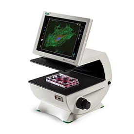
Take cell imaging out of the darkroom and into your classroom! The ZOE Fluorescent Cell Imager is a complete digital imaging system, allowing students to view samples, capture and store images, and create multicolor overlays.
The 10.1-inch screen and HDMI projector connectivity allows all your students to view live images simultaneously. With a compact 13 x 12.6 inch footprint, the ZOE Cell Imager is easily transportable from your teaching lab to your classroom.
Features and Benefits of the ZOE Fluorescent Cell Imager
- Fluorescence imaging in your classroom — light shield permits fluorescence imaging in ambient light and eliminates the need for a darkroom
- Simplified cell imaging — the intuitive touch-screen interface allows students to view cells, capture images, and create multichannel merges with minimal training
- HDMI connection — project live microscopy images, allowing the entire class (including online students) to see the same thing at the same time
- Flexible operation — brightfield and three fluorescence channels enable your students to learn both simple and sophisticated imaging techniques
- Robust construction — fully integrated system with long-life LEDs, ready for the wear and tear of teaching duty
- Small footprint — compact size accommodates crowded lab benches and easy transportation
Applications of the ZOE Fluorescent Cell Imager
With the ZOE Cell Imager’s brightfield and three fluorescent channels, your students can experience cell culture work and learn fluorescent techniques practiced regularly in the research lab:
- Observation of general cell health and morphology
- Monitoring cell growth and proliferation
- Estimation of transfection efficiency
- Visualization of expressed fluorescent proteins
- Immunofluorescent protein localization
- Visual estimation of cell confluency
- Capturing cell images (with or without fluorescent labels)
You can use the ZOE Cell Imager with various dyes and fluorophores to teach your students about cellular biology, gene expression, protein localization, developmental biology and genetics, morphology, and much more.
Specifications
Specifications
Green channel: blue LED
Red channel: green LED
Brightfield channel: multiple green LEDs (reduces chromatic aberration)
Multiwell plates: 6-, 12-, 24-, 48-, 96-, or 384-well microplates
Dishes: 35 mm, 60 mm, or 100 mm
Slides: chamber slides or standard glass microscopy slides
