
This section provides information on nucleic acid electrophoresis. The different types of electrophoresis cells, agaroses, and reagents available for this technique are presented and discussed.
Related Topics: Nucleic Acid Extraction and Purification.
Page Contents
Gel electrophoresis is an analytical technique used to separate nucleic acid molecules (DNA or RNA) based on size. This technique can effectively separate molecules up to ~20kb in size. Nucleic acid separation by gel electrophoresis has numerous applications for analysis in molecular biology, following PCR analysis or before cloning, sequencing, northern or Southern blotting.
Nucleic acids that are to be analyzed are dispensed into wells of an agarose gel. The agarose gel acts as a matrix to contain and separate the target molecules. The gel is submerged in an electrophoresis chamber with buffer that allows the flow of an electric current. An electromotive force is applied across the chamber. Buffer solutions act to reduce pH changes due to the electric field and to prevent any overheating of the gel that may be caused by an electric current.
For accurate estimation of the fragment size, nucleic acid standards with bands of known size are loaded in the gel next to actual samples. These commercially available products have evenly spaced banding patterns and are available in different size ranges.
Nucleic acid molecules are colorless and require staining for visualizing. A tracking dye such as bromophenol blue is often used for easy monitoring of their movement in the gel. Ethidium bromide is a sensitive fluorescent stain for visualizing DNA in agarose and polyacrylamide gels. It is commercially available in a ready-to-use solution for convenience. Stained gels are visualized using a gel imaging system such as the Gel Doc EZ System.
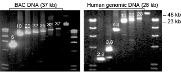
Nucleic acid gel electrophoresis.
Electrophoresis gel boxes are an integral part of nucleic acid separation. A gel box is a container that can accommodate a gel tray and has electrodes that can be connected to a power supply. The electrodes on the gel box are usually color coded: black for the cathode and red for the anode. The cathode carries the negative charge while the anode carries the positive charge. When the gel box is connected to a power supply, electrons enter through the cathode and leave through the anode. The flow of electrons sets up a potential energy difference between the electrodes and establishes an electric current. The gel box chamber holds the gel and is filled with a buffer prior to passing any electric current.
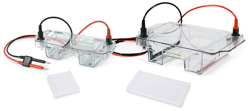
Horizontal electrophoresis gel boxes.
Gel Box Assembly
Gel boxes are either horizontal or vertical in format. Horizontal gel boxes are the common choice for separating nucleic acid fragments, while vertical gel boxes are more commonly used for protein electrophoresis.
Gel boxes can come in varying sizes, and choosing the right gel box depends on a couple of factors: the number of samples to be analyzed and the molecular weight of the DNA fragments to be separated. A wider chamber can accommodate more samples in parallel, while a longer gel box enables placing samples in multiple rows or can be used to run samples for a longer distance through the gel. Larger formats are ideal for restriction fragment length polymorphism (RFLP), Southern blotting, and multiple sample screening. Mini formats are useful for quick separations and smaller numbers of samples.
Bio-Rad provides many options for gel boxes in varying widths and lengths.
 Mini-Sub Cell GT* |
 Wide Mini-Sub Cell GT** |
 Sub-Cell GT |
 Sub-Cell Model 96 |
 Sub-Cell Model 192 |
|
| Cell size (W x L x H) |
9.2 x 25.5 x 5.6 cm | 17.8 x 25.5 x 6.8 cm | 18 x 40.5 x 9.4 cm | 29 x 30 x 9 cm | 29 x 40 x 9 cm |
| Gel tray sizes (OD) (W x L) |
7 x 7 cm 7 x 10 cm |
15 x 7 cm 15 x 10 cm |
15 x 10 cm 15 x 15 cm 15 x 20 cm 15 x 25 cm |
25 x 10 cm 25 x 15 cm |
25 x 10 cm 25 x 15 cm 25 x 20 cm 25 x 25 cm |
| ReadyAgarose gels accommodated | Yes | Yes | No | No | No |
| Sample throughput | 8–30 | 10–60 | 1–120 | 24–96 | 24–192 |
| Base buffer volume | ~270 ml | ~650 ml | ~1 L | ~2 L | ~3 L |
| Buffer recirculation | No | No | No | Yes | Yes |
Agarose is a linear polysaccharide composed of alternating residues of D- and L-galactose that are joined by glycosidic linkages. Agarose gels are made by dissolving agarose in buffer to form a porous and resilient matrix for nucleic acid separation. This forms a sieving matrix which allows the electrophoretic separation of charged macromolecules, such as DNA or RNA, according to size.
There are various agarose grades that are commercially available.
Molecular biology grade: This general purpose agarose is used for most routine purposes of nucleic acid separation. For most conditions this agarose offers rapid migration rates and easy-to-manipulate gels.
PCR grade agarose: This agarose grade is recommended for separation of PCR fragments. It is especially useful for fragments smaller than 1 kb.
Low range ultra agarose: This grade provides superior resolution for smaller fragments, 10-100 bp in size, and can resolve differences of ~5 bp.
Low-melt agarose is also occasionally used for specific applications involving DNA and RNA recovery.
Agarose gels can be prepared in the laboratory (handcast) or purchased premade (precast) from many vendors.
- Handcast agarose gels — agarose gels are prepared by mixing agarose in either TAE or TBE buffer by weight/volume measurements to get the desired percentage of agarose consistency. Gels are described in terms of percents: 0.8%, 1%, 1.2% etc. The percentage gel chosen for an experiment depends on expected fragment size and desired separation of fragments.
- Precast agarose gels — numerous precast gels are available commercially. These are designed to fit securely in compatible gel boxes. ReadyAgarose™ precast gels from Bio-Rad come in many sizes and agarose percentages and can resolve nucleic acids ranging from 20 to 20,000bp.
Buffers commonly used for DNA agarose electrophoresis are either TAE (Tris-acetate with EDTA, 40mM Tris-acetate, 1mM EDTA) or TBE (Tris-borate with EDTA, 89mM Tris-borate, 2mM EDTA). The neutral pH of these buffers is critical, as it allows the phosphate backbone of DNA that has a net negative charge to migrate toward the anode. TAE and TBE have different properties, so one is more suitable than the other for any specific application. TAE buffer has relatively low buffering capacity and is recommended for electrophoresis of larger DNA fragments (>12 kb). TBE has higher buffering capacity, which decreases DNA mobility and is recommended for electrophoresis of small DNA fragments (< 1kb).
While both TAE and TBE are relatively simple to prepare, they are also commercially available. Bio-Rad provides premixed nucleic acid electrophoresis buffers that are ready to be used.
Nucleic acid buffers and reagents.
Sambrook J et al. (2001). Molecular Cloning: A Laboratory Manual (Cold Spring Harbor, New York: Cold Spring Harbor Laboratory Press).
Singh SP et al. (1999). Use of pulsed-field gel electrophoresis for molecular epidemiologic and population genetic studies of Mycobacterium tuberculosis. J Clin Microbiol 37, 1927–1931. PMID: 10325348